Scanning Electron Microscope Service
Scanning electron microscope (SEM) is an observation method. It does not use electromagnetic lens to amplify the image but uses a narrow focused-high-energy electron beam to scan the sample, which in a similar way of television photography. SEM stimulates various physical information through the interaction between the beam and the material, and then the sample information will be collected, enlarged, and reimagined to achieve the purpose of characterizing the micromorphology of the material.
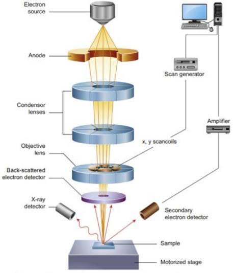 Figure 1. Schematic diagram of the core components of an SEM (Inkson B J. 2016)
Creative Biostructure can provide SEM service for our customers. SEM has been widely used in biology, archaeology, petroleum exploration, chemistry, medicine, justice, material sciences and other fields. With high-resolution electrons, SEM can be used to observe the sample surface, element analysis, crystal structure analysis, two-dimensional morphology observation and analysis. Moreover, except for displaying the morphology of the general sample surface, it can also display the differences between the chemical elements, the properties of light, electricity, and magnetism of the sample micro area. In addition, with wide application range and simple operation of SEM, various types of materials can be observed by SEM. For example, cells, tissues, blocks, films, powders and even biopolymers can be directly tested with little or no treatment.
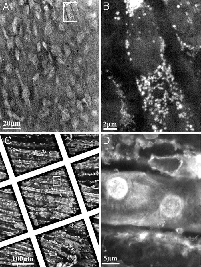 Figure 2. SEM Imaging of tissues (Stephan T, et al. 2004)
We can also provide cryo-SEM services. Using rapidly freezing methods, cryo-SEM had been widely used in cell samples due to it preserves the natural structures of the sample. The conventional SEM method will dry the materials during the sample preparation process, which may affect the sample morphology, especially the biological sample of high-water content. While the freezing technology can avoid cell deformation and has the advantages of simple and fast sample preparation. In addition, compared with the environmental scanning electron microscope (SEM), cryo-SEM can directly observe water samples, and freezing scanning can be observed under high vacuum conditions, which improves the resolution.
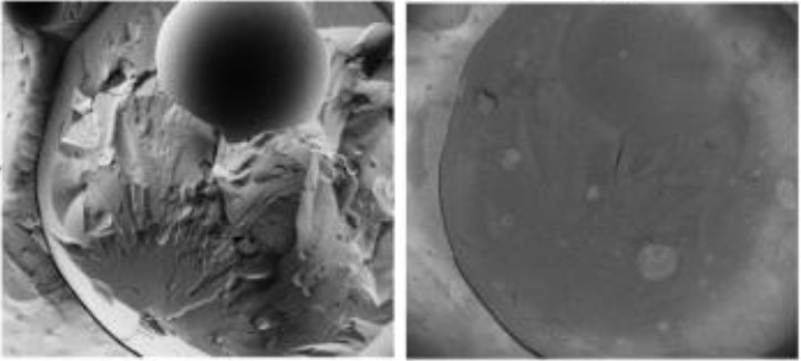 Figure 3. Cryo-SEM of high internal phase multiple O/W/O/W emulsion (Lucy L, et al. 2020)
Creative Biostructure will process different samples according to their characteristics, and finally complete the data collection and analysis.
If you are interested in our scanning electron microscope services, please feel free to contact us. We are looking forward to cooperating with you.
Ordering Process

References
- Inkson B J. Scanning electron microscopy (SEM) and transmission electron microscopy (TEM) for materials characterization. Materials Characterization Using Nondestructive Evaluation (NDE) Methods. 2016:17-43.
- Stephan T, et al. Scanning electron microscopy of cells and tissues under fully hydrated conditions. Proc Natl Acad Sci. 2004, 101(10): 3346-3351.
- Lucy L, et al. Micrograph contrast in low-voltage SEM and cryo-SEM. Ultramicroscopy. 2020, 218: 113085.

