In a recent study, scientists took the first overall color picture of mice of different ages, which is an important step in understanding individual behavior. The results of the study, published in the journal Science, are key to revealing the mechanisms of learning disabilities, dementia, and how memory is affected by age.
Synapses are important connections between brain cells to transmit electrical and chemical information. Synapse damage is associated with more than 130 brain diseases. In this study, researchers at the University of Edinburgh color-coded different types of molecules to highlight the range of synapses in the brains of mice of different ages from birth to old age.
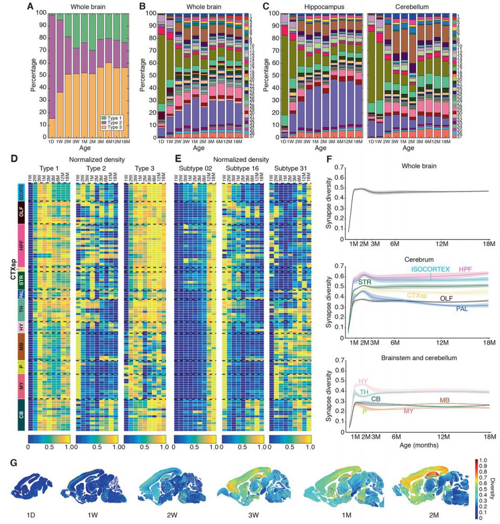
They found that in different parts of the brain, the number and molecular composition of synapses change with age. These changes occur in three main stages, children, middle age, and old age.
The type of synapse changes with age, which is a unique pattern in the brain area, and develops into various forms in middle age. The images from the middle-aged brain are complex, indicating that the diversity of synapses is higher at this time. In contrast, the brains of young and old mice show fewer synapses and less complexity.
The researchers say these changes can reveal why it is easy to cause synaptic damage at certain ages and in certain brain regions. At the same time, these findings can clarify why we are more susceptible to brain diseases at certain ages. For example, why schizophrenia usually starts in adolescence, and dementia mainly affects the elderly.
Reference
“A brain-wide atlas of synapses across the mouse lifespan” Science (2020). URL:science.sciencemag.org/lookup/ … 1126/science.aba3163