According to a recently published report in Nature Communications, scientists from Duke University Medical Center and other institutions revealed how stem cell mutations quietly occur and spread to a wide range of areas in the colon until they eventually dominate and develop into malignant tumors. By using an innovative model system in mice, researchers can visually mark colon cancer mutations by promoting stem cell luminescence, and then they can observe mutations that occur in colon cancer in animals and shed light on the intestinal tract. And it can clarify a process from death to death that occurs in the intestine. One mutation will overcome the other mutation and eventually become the driving force for malignant tumors.
Researcher Joshua Snyder said that this study reveals a process we have not observed before: how mutant precancerous stem cells spread in the colon and sow the seeds of cancer. The technology we use lays a solid foundation for testing new therapies, and these therapies will eventually effectively block early precancerous processes; researchers hope that one day they can target and clear these precancerous cells to effectively prevent cancer.
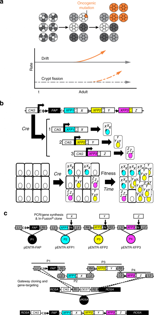
In the article, the researchers used a molecular staining technique that can mark multiple common colon cancer mutations in stem cells from a single tumor, resulting in a fluorescent barcode. When transferred to mice, the fluorescent stem cells can be effectively tracked, revealing the cellular and molecular dynamics of precancerous events. Researchers have found a key difference in the generation of precancerous mutant cells in the intestinal dwelling tissues of infants and adults. At critical times, newborns are particularly sensitive to the mutational effects of intestinal stem cells, which will unknowingly seed a large number of precancerous mutations in their intestines (a process known as regional canceration), thereby increasing the risk of neonatal disease. These mutated cells will grow and spread, which cannot be detected by current screening techniques. Of course, these cells are generally harmless, but if appropriate, they can develop into cancer soon after adulthood.
Researchers have pointed out that certain colon cancer mutations in the patient’s body can cause an increase in “fertility” in the surrounding environment of the precancerous area, eventually leading to the rapid spread of cells in the intestine and causing fatal consequences. Certain common mutations caused by external sources (such as injury or environmental exposure) can interfere with the environment surrounding stem cells and cause rapid growth and spread of cells in the precancerous area. These conditions are particularly deadly to adults and occur much faster than previously expected, as if throwing matches in an arid forest.
Researcher Snyder said that field cancerization is considered to be the decisive event initiating the growth process of cancer, including breast cancer, skin cancer, and lung cancer. How cells compete and expand in a field, which promises to help develop technologies and novel therapies for early diagnosis. Researchers are currently conducting other studies to use fluorescent barcodes to observe cancerous fields in breast cancer. They are aiming to clarify whether precancerous lesions such as ductal carcinoma in situ are caused by malignant or benign mutations.
Reference
Peter G. Boone, Lauren K. Rochelle, Joshua D. Ginzel, et al. A cancer rainbow mouse for visualizing the functional genomics of oncogenic clonal expansion. Nature Communications, 2019; 10 (1) DOI: 10.1038/s41467-019-13330-y