On the Feb. 10th, 2018, it was learned from the University of Science and Technology of China that researchers at the university made breakthroughs in resolving the ultrastructure of synapses using cryo-electron microscopy and decrypting the black box[1] of synapses.
Recently, The Journal of Neuroscience, an international journal of the American Academy of Neuroscience reported this discovery in the form of a journal cover.
Synapse is the most basic structure and functional unit of brain function, consciousness, learning, and memory, and is also the origin of many brain diseases. It is believed that accurately resolving the molecular structure of synapses and their changes during neural activity is the most direct and effective way to decrypt the brain’s mysteries. It is also one of the most basic research work in neuroscience. Early, biochemical and molecular biology, electrophysiology and other research found a variety of large molecules and organelles in synapses and revealed its various functional characteristics and plasticity rules. However, due to limitations of research methods, how these different components of the synapse organize into complex machines to perform different functions has not been adequately observed and resolved.
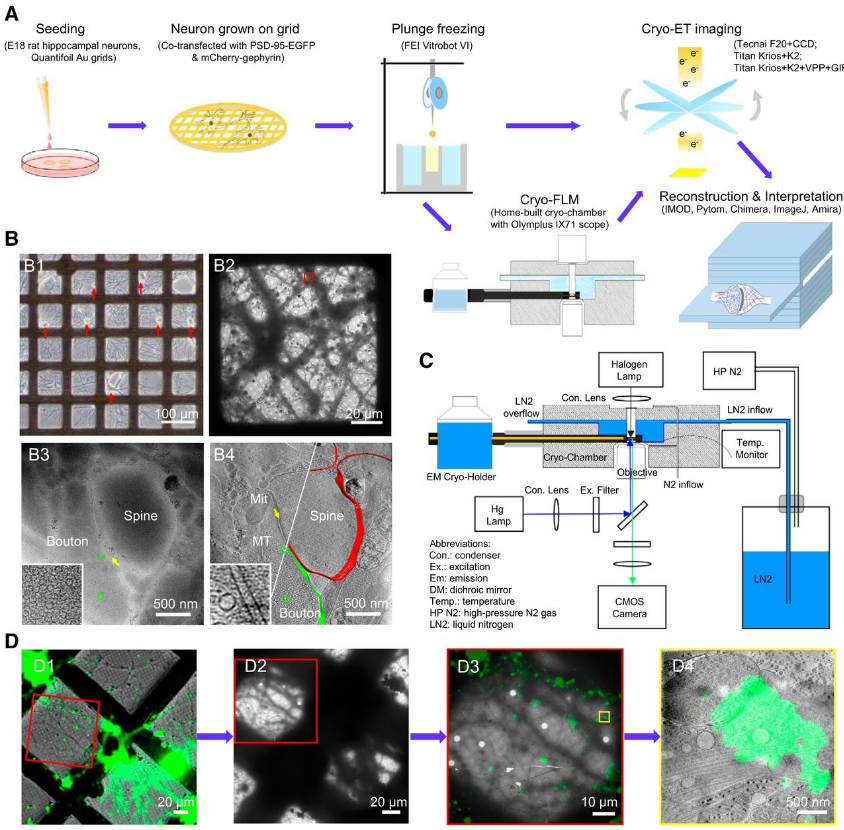
Professor Bi Guoqiang, from National Laboratory for Physical Sciences at the Microscale, and professor Zhou Zhenghong, Liu Beiming from Chinese Academy of Sciences Key Laboratory of Brain Function and Disease, School of Life Sciences, cooperated and achieved the quantitative analysis of the two main types of synapses in the CNS using the latest development of CryoET three-dimensional reconstruction technology (cryoET), combined with self-developed frozen Photoelectric Correlation Microscopy. The team obtained a series of three-dimensional structures of intact synapses in the almost physiological state by culturing rat hippocampal neurons on the special network of cryo-microscopy.
With the quantitative analysis method, it was the first time to report inhibitory synaptic uniform laminar post-synaptic structure and found ellipsoidal synaptic vesicles existing in two types of synapses. And it brought the ending to the long-standing controversy over the fine structure of both synaptic vesicles and postsynaptic densities.
Subsequently, the team further obtained the fine structure of the synapse at the molecular level and achieve a direct observation in the interaction between single neurotransmitter receptor protein complex and its scaffold proteins in situ in synapses.
This is the first systematic and quantitative analysis of intact synapses by cryo-microscopy in the world for the first time. This discovery, on one hand, promoted the decryption of the “black box” of synaptic ultrastructure and function, on the other hand, it lays a foundation for breaking the technical problem of cryogenic microscopy in situ analysis of the structure of biomacromolecule complex in a complex cell system.
Get more information on cryoET service on our website.
(With the combination of 3D imaging and specimen preparation that preserves the integrity of the cell structure, Cryo-ET can observe the internal structure of the cell with high resolution.)
[1] Black Box: The working mechanism, which had not be discovered before.
Reference
Chang-Lu Tao, et al. Differentiation and Characterization of Excitatory and Inhibitory Synapses by Cryo-electron Tomography and Correlative Microscopy.
Wow, thiѕ article is nice, my sister is analyzing these kinds
of things, therefore I am going to inform һer.