MagHelix™ Scanning Electron Microscopy (SEM) Products
Creative Biostructure has abundant experience in using MagHelix™ SEM to reveal the surface topography and composition of target samples. A large variety of high quality products related to SEM specimen preparation are available.
| Products | Descriptions | Categories |
| Conjugates | Labeling of proteins for electron microscopy detection. |
Gold conjugate Silver enhancing kits Blocking reagents |
| Fixation chemicals | Dehydrating of specimen for embedding |
Glutaraldehyde Osmium tetroxide Gradient ethanol |
| Mounting adhesives | Mounting of specimens permanently or temporarily for viewing |
Conductive gold DAG Conductive graphite paint Specimen bonding adhesives Fast drying silver suspension Epoxy Resin |
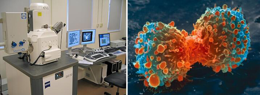
SEM is a type of Electron Microscope(EM) that generates living images of a biological sample by scanning across the surface of the sample with a focused electron beam. The most common SEM is detection of secondary electrons emitted by atoms excited by the electron beam.
All samples prepared for SEM must be of an appropriate size to suit chamber and are generally mounted rigidly on a specimen holder. Due to the SEM uses vacuum conditions, special preparations must be done to the sample. For instance, first, all water must be removed from the samples because the water would vaporize in the vacuum; then, all metals are conductive and require no preparation before being utilized; finally, all non-metals need to be made conductive by covering the sample with a thin layer of conductive material.
Creative Biostructure aims at providing fast and efficient MagHelix™ Electron Microscopy (EM) services to accelerate customers’ researches and projects, and is ready to be of assistance 24/7. We can also offers various custom UniCrys™ membrane protein crystallization services and products for TEM and cryo-EM. Please feel free to contact us for detailed information.
References:
L. Reimer (1998). Scanning electron microscopy: physics of image formation and microanalysis. Springer, 527 p.
Scanning electron microscope. (https://en.wikipedia.org/wiki/Scanning_electron_microscope).