As we all know, an optical microscope for direct observation is of great significance in biomedical research, such as cell biology, developmental biology, immunology, pathopharmacology, and so on. However, due to the limitation of the diffraction limit, the resolution of the traditional optical microscope can only reach half of the light wavelength theoretically. For a long time, the appearance of super-resolution fluorescence microscopy has effectively broken the limit of optical diffraction. Super-resolution microscope (SMLM), stimulated emission loss microscope (STED) and structured light illumination super-resolution microscope (SIM) based on single-molecule localization technology have made great progress with the efforts of many research groups. Especially, a structured light illumination microscope has the advantages of fast imaging speed, low phototoxicity, and no need for special fluorescent labeling. It has become a popular technique in the field of life science, especially in the imaging of living cells. Recently, the Li Hui research group of Suzhou Institute of biomedical engineering technology, Chinese Academy of Sciences has made a series of important progress in the research of structured light illumination super-resolution microscopy imaging method, high fidelity SIM reconstruction algorithm, and the development of domestic SIM microscope.
The three-dimensional imaging method has attracted much attention because it can obtain more information about biological samples. However, the existing three-dimensional imaging inevitably brings the problems of defocus blur and poor time resolution, which is not easy to be used for fast three-dimensional dynamic imaging of samples. In order to realize fast three-dimensional imaging of thick samples, Li Hui’s research group has developed a structured light illumination slicing microscopy technology based on digital micromirror array device (DMD) and liquid zoom lens (ETL), and developed a slicing imaging algorithm based on two original images. This method improves the speed of traditional three-dimensional slice imaging by several times. Using this technology, the research group has carried out high-speed dynamic imaging of the cardiovascular system of zebrafish and cerebral vessels, which clearly shows the systolic diastolic process of the heart beating period and the peristaltic characteristics of abdominal vessels. The related research achievements are entitled four-dimensional visualization of zebrafish cardiovascular and vessel dynamics by a structured illumination microscope with electrically tunable lens and published in Biomedical Optical Express (2020).
Structured light illumination super-resolution imaging technology has been widely used in a variety of nanoscale subcellular structure research. However, for samples with a large dynamic range (such as aggregated cell vesicles), the aggregated regions with strong fluorescence and sparse regions with weak brightness cannot be presented at the same time. The existing SIM methods can’t reconstruct high-quality images for this kind of sample. In this regard, Li Hui’s research group proposed a high dynamic SIM imaging method HDR-SIM, which uses multiple exposure acquisition. Three groups of SIM images with different illumination intensities are collected, and then a super-resolution image is fused. Using HDR-SIM, the intensity difference is more than 400 times, and the single and aggregated fluorescent beads can be observed simultaneously in the same SIM super-resolution image, and the resolution will not be affected. When using this method to observe the structure of cell vesicles of different scales, both single small vesicle and large vesicle aggregation can be distinguished clearly at the same time. The related research results are published on Frontiers in Physics (2021) with the title of high dynamic range structured illumination microscope based on multiple exposures.
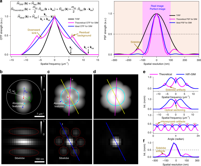
In the process of structured light illumination imaging, a super-resolution image reconstruction algorithm is particularly critical. Due to some inherent defects of the SIM reconstruction algorithm, reconstruction artifacts often appear in super-resolution images, so the fidelity of SIM images is often questioned, and a series of complex parameter settings need to be completed in image reconstruction, which limits the application of SIM technology to ordinary users. Li Hui’s research group has developed a high-fidelity SIM reconstruction algorithm based on point spectrum optimization. This algorithm overcomes the problems that the conventional SIM algorithm is easy to produce reconstruction artifacts and poor optical slicing ability. High-quality super-resolution images with few artifacts and good optical slicing can be obtained by processing different quality raw data, which effectively improves the fidelity of SIM imaging. At the same time, the algorithm is not sensitive to OTF mismatch and user-defined parameters. High-quality SIM images can be reconstructed by using the generated theoretical OTF and fewer parameters, which reduces the high requirements of SIM imaging for experimental implementation and post-processing reconstruction, and improves the friendliness of the algorithm to ordinary users. Compared with several traditional SIM algorithms, the HIFI-SIM algorithm reconstructs a variety of original data with different image quality, different sample complexity, and different image sources (commercial equipment / self-built SIM system). The HIFI-SIM algorithm shows the least reconstruction artifacts and the best image quality. The related research results are published on light: Science & Applications (2021) with the title of high fidelity structured illumination microscope by point spread function engineering.