Scanning Tunneling Microscopy (STM) Service
Scanning Tunneling Microscope, STM, was developed in 1982 by a research group led by G. Binnig and H. Rohrer in IBM Zurich Laboratory. The advent of STM has fulfilled the centuries-old dream of scientists to observe atoms and molecules directly, greatly promoting the development of surface science and nanoscience.
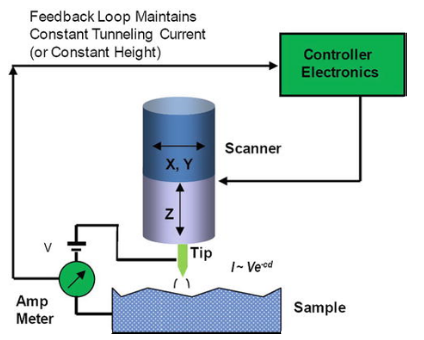 Figure 1. Schematic of the scanning tunneling microscope. The tip is shown mounted on a piezoelectric tripod with three orthogonal scanners marked x, y, and z. A bias is applied between the tip and the substrate. The feedback loop maintains either constant-current or constant-height operation mode.
STM makes use of quantum theory's tunneling effect, the quantum behavior of microscopic particles such as electrons that can penetrate or pass through a potential barrier, even though the barrier is higher than the total energy of the particle. The core component of STM is the tip of the probe. The basic principle of STM is to use the very thin probe of atomic alignment and the surface of the substance under study as two electrodes. When the distance between the sample and the tip is very close (usually less than 1nm), under the action of an applied electric field, electrons will flow through the potential barrier between the two electrodes to the other electrode. The current generated at this time is called tunnel current. By scanning and recording the different tunnel currents at each point on the surface, the morphology of the sample surface can be described, as shown in the figure.
STM has two operating modes, which are constant-height mode (keep the height of probe tip and sample unchanged, measure the change of current to depict the morphological characteristics) and constant-current mode (keep the current constant, depict the morphological characteristics through the change of measurement height).
STM applications:
- Due to the unique shape and principle of the tip, it has a very high resolution, which is usually used to scan the morphological characteristics of samples.
- Using STM to move atoms can perform microscopic precise operations.
- Spectrum imaging measurement can be performed.
Creative Biostructure has a lab dedicated to protein structure analysis with many years of experience. We have optimized the STM techniques during several years of working. Our STM is capable of multi-temperature testing compared to ordinary STM technology and is gradually supporting multi-substance sample testing.
Creative Biostructure provides STM booking and image analysis services. We can provide you with common tomograms, atomic resolution maps, and common density of states.
If you are interested in our STM service, please feel free to contact us. Creative Biostructure looks forward to cooperating with you.
Ordering Process

Reference
- He Y., Chen S., Yu Q. (2013) Scanning Tunneling Microscope (STM). In: Wang Q.J., Chung YW. (eds) Encyclopedia of Tribology. Springer, Boston, MA. https://doi.org/10.1007/978-0-387-92897-5_1225


