Single Particle Cryo-EM Data Analysis for Coronavirus Research
The three-dimensional (3D) reconstruction technology of single-particle cryo-electron microscopy (cryo-EM) involves a complex image processing process from two-dimensional (2D) projection images to a 3D reconstruction model. Creative Biostructure currently provides specialized services about single-particle cryo-EM data acquisition and 3D reconstruction of biological macromolecular to promote the progress of coronavirus infection research.
Brief Introduction to Coronavirus and SARS-CoV-2
Novel coronavirus (also known as SARS-CoV-2) genome-wide sequence analysis showed that it belongs to β-coronavirus, but is different from the severe acute respiratory syndrome coronavirus (SARS-CoV) and Middle East respiratory syndrome coronavirus (MERS-CoV). Together with Bat SARS-like coronavirus, SARS-CoV-2 forms a unique lineage within the subgenus of Sarbecovirus. There is currently no specific prevention and treatment method.
Recently, the full-length 3D structure of the SARS-CoV-2 host cell receptor ACE2 has been successfully analyzed through single-particle cryo-EM technology, and it was found that ACE2 existed as a dimer. In addition, the structure of the receptor-binding domain (RBD) of the SARS-CoV-2 spike protein with full-length ACE2 complex has also been successfully resolved, which can provide a good reference for understanding the interaction between the two and the subsequent design of targeted antiviral drugs.
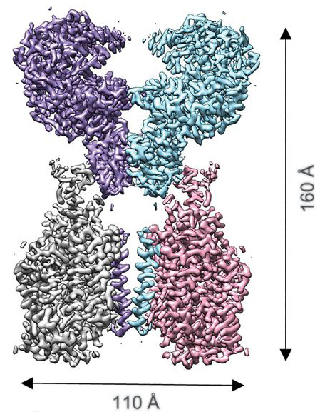 Figure 1. Cryo-EM map of the ACE2-B0AT1 complex. (Adapted from Yan R.; et al. 2020)
Cryo-EM Data Acquisition and Analysis for Coronavirus Research
- Sample screening and data acquisition
After preparing a frozen-hydrated sample of biological macromolecules, we will evaluate the sample and select the sample with the optimal particle density and thickness of glassy ice that is structurally uniform and are most likely to generate the best image. We utilize a highly stable 300kV transmission electron microscope for large-scale data acquisition. In this process, we set the optimal parameters (such as underfocus values, magnification, and the total electron dose, etc.), and capture and record a large number of images of these sample areas.
- Data processing and analysis
Creative Biostructure can provide cryo-EM image processing, 3D reconstruction, and structural analysis services. We use manual or automatic programs to select projection images, align and merge the data through various image processing methods and professional image processing software, and then build the initial 3D model. The further iterative process allows for refining and verifying the model, and then the 3D structure model of biological macromolecules can be reconstructed, and finally the structure analysis and evaluation can be carried out.
Why Do You Choose Us?
- First-class electron microscopy platform and experienced team of structural biology experts
- Our scientists have extensive knowledge in the field of virology research
- Flexible services, biomacromolecule samples for cryo-EM can be provided by our customers or prepared from our high-purity protein expression and purification services
- We are strictly confidential about project information, experimental data, and analysis results
- Our customer service representatives are available 24 hours a day from Monday to Sunday
Contact us to discuss your project!
References
- Yan R; et al. Structural basis for the recognition of SARS-CoV-2 by full-length human ACE2. Science. 2020, 367(6485): 1444-1448.
- Wrapp D.; et al. Cryo-EM structure of the 2019-nCoV spike in the prefusion conformation. Science. 2020, 367(6483): 1260-1263.
- Doerr A. Single-particle cryo-electron microscopy. Nature Methods. 2015, 13(1): 23.