Super-resolution Microscopy for Coronavirus-like Particle
Because most viruses are small (< 200 nm), standard optical microscopes are generally not suitable for direct observation of them. The use of electron microscopy (EM) to visualize the ultrastructure of viruses has become the gold standard for viral imaging, however, the complexity of preparing biological samples (fixed or frozen) and the need to place specimens under vacuum make it unsuitable for studying the dynamic processes during virus-cell interactions. Creative Biostructure is now capable of providing super-resolution imaging services using various super-resolution microscopes for your coronavirus-related research. Super-resolution microscopes can be used to observe biological three-dimensional (3D) structures that many conventional fluorescence microscopes cannot resolve, and to measure interactions and record dynamic processes in living cells at the nanometer level.
Application of Super-resolution Microscopy in Virology
With the development of super-resolution fluorescence microscopy (SRFM) and nanotechnology, the impasse in viral imaging technology has changed dramatically around the diffraction limit of viruses to improve resolution. These techniques of super-resolution microscopes can routinely provide a spatial resolution of 10-100 nm and can even achieve a resolution as low as 1 nm. Super-resolution microscopy technologies combine the higher resolution that can solve subviral structures with all the advantages of fluorescence microscopy. Due to the advantages of non-invasive living cell imaging, labeling specificity, and high-throughput, SRFM is an ideal tool for in-depth studies of virus substructures and virus-cell interactions. To date, only a few reports have used this technology to study the biology of viruses. Among them, SRFM has provided many novel insights into the replication cycle of retroviruses, particularly human immunodeficiency virus type 1 (HIV-1).
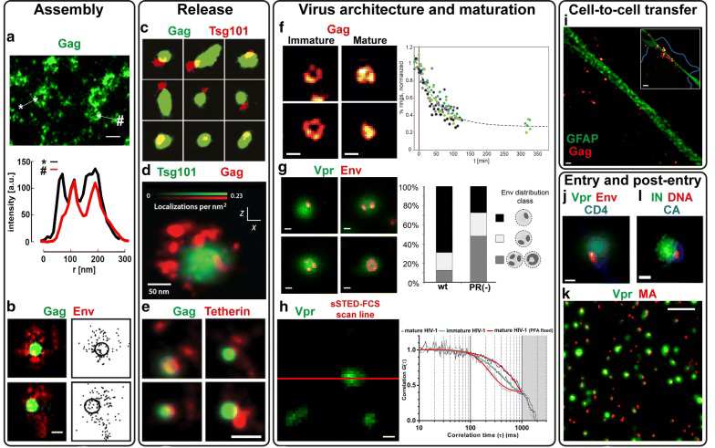 Figure 1. SRFM studies on the HIV-1 replication cycle. (Adapted from Chojnacki J, Eggeling C. 2018)
Our Super-resolution Microscopy Services for Coronavirus-like Particle
Currently, a variety of SRFM techniques have been developed, and we support several far-field imaging methods that can extend the diffraction-limited resolution to smaller values, such as stimulated emission depletion (STED) microscopy, (direct) stochastic optical reconstruction microscopy [(d)STORM] and photoactivation localization microscopy [(f)PALM]. You can obtain the following information through our super-resolution microscopy services:
- 3D viral imaging and multicolor imaging
- Dynamic behavior of individual virus substructures
- Interaction of viruses and cellular components in living cells or tissues
For public safety considerations, we can only accept coronavirus-related samples without high infectivity, and the service is not used for diagnostic tests but only for research purposes. If you are interested in our super-resolution microscopy services for coronavirus-like particles, please do not hesitate to contact us. And our customer service representatives are available 24 hours a day from Monday to Sunday.
Contact us to discuss your project!
References
- Grove J. Super-resolution microscopy: a virus’ eye view of the cell. Viruses. 2014, 6(3): 1365-1378.
- Chojnacki J, Eggeling C. Super-resolution fluorescence microscopy studies of human immunodeficiency virus. Retrovirology. 2018, 15(1): 41.