Single Particle Cryo-EM Sample Preparation for Coronavirus Research
The latest technological advances in sample preparation, algorithm, and detector have made single-particle cryo-electron microscopy (cryo-EM) analysis a popular approach for solving macromolecular structures at near-atomic-resolution. After obtaining high-purity coronavirus-associated protein of interest, Creative Biostructure provides cryo-EM sample preparation and vitrification services to support cryo-EM observation and data collection.
Brief Introduction to Coronavirus and SARS-CoV-2
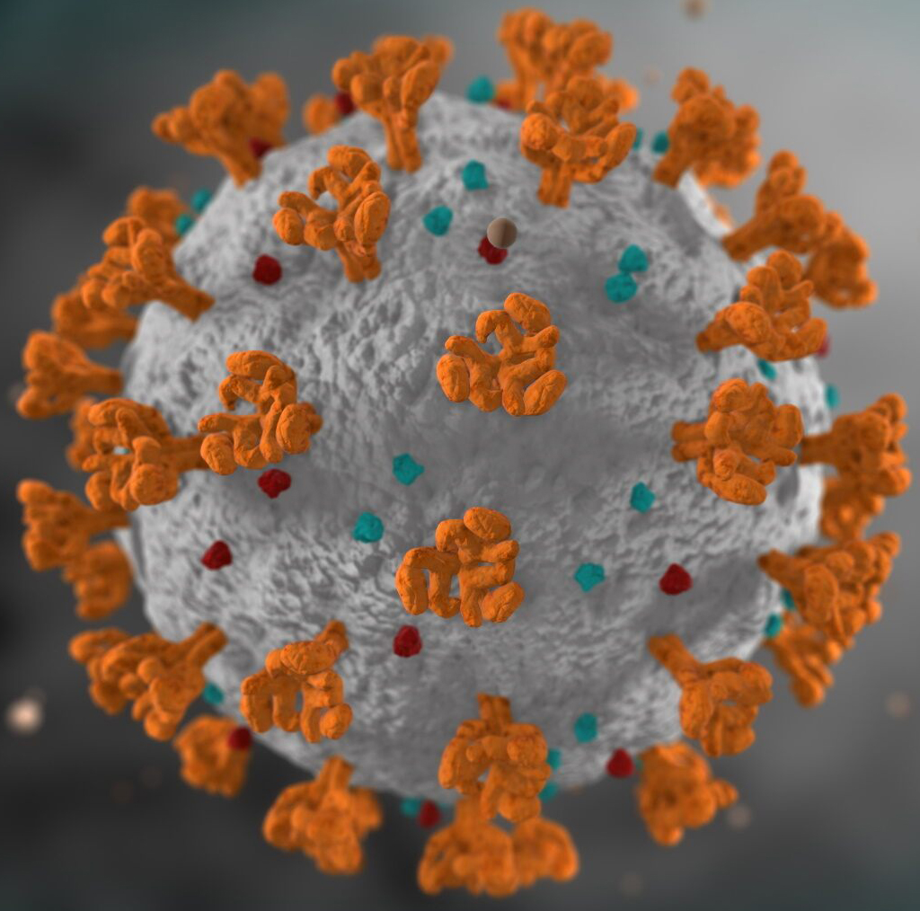
The novel coronavirus (also known as SARS-CoV-2, formerly 2019-nCoV) outbreak has been declared as a public health emergency of international concern. The spike glycoprotein (S protein) of novel coronavirus has also become a crucial target for the development of diagnostic methods, vaccines, and therapeutic antibodies. At present, scientists have successfully analyzed the full-length three-dimensional (3D) structure of the SARS-CoV-2 cell surface receptor ACE2 and the 3D structure of the S protein receptor-binding domain (RBD) in complex with ACE2 using single-particle cryo-EM technology. The analysis of these cryo-EM structures provides an important structural biology foundation for further accurate vaccine design and discovery of antiviral drugs.
Cryo-EM Sample Preparation and Vitrification for Coronavirus Research
The cryo-EM experiment starts with purified protein samples. High-purity coronavirus-associated protein samples can be provided by our customers or prepared through our protein expression and purification service. Usually a small amount of protein solution can be used to prepare cryo-EM samples. The protein solution is applied to a metal grid with an additional layer of continuous thin carbon films. Ideally, the protein particles are evenly distributed in the grid holes in various orientations. For compatibility with the high vacuum conditions of the transmission electron microscope (TEM) and to lock the single particle in its natural state, we will vitrify the sample. The grid adsorbed with the protein solution is then plunged into the cryogen (liquid ethane), flash-freezing it and trapping the protein particles in a thin film of vitreous ice. Baked by our state-of-the-art equipment, high-quality vitrified samples can be reproducibly prepared.
Why Do You Choose Us?
- Professional structural biology contract research provider with advanced facilities
- Drawing on the advanced experience in the industry, constantly optimizing the technology of cryo-EM sample preparation
- Services are not bundled, and competitive prices for multiple service needs
- High transparency, timely communication and feedback
- Our customer service representatives are available 24 hours a day from Monday to Sunday
Contact us to discuss your project!
References
- Yan R; et al. Structural basis for the recognition of SARS-CoV-2 by full-length human ACE2. Science. 2020, 367(6485): 1444-1448.
- Wrapp D.; et al. Cryo-EM structure of the 2019-nCoV spike in the prefusion conformation. Science. 2020, 367(6483): 1260-1263.
- Doerr A. Single-particle cryo-electron microscopy. Nature Methods. 2015, 13(1): 23.