MagHelix™ Transmission Electron Microscopy (TEM) Products
Scientists from Creative Biostructure have extensive experience in specimen preparation for TEM. And we also provide various products for the preparation procedures.
| Products | Brief description | Categories |
| Conjugates | Labeling of proteins for electron microscopy detection. |
Gold conjugate
Silver enhancing kits Blocking reagents |
| Wax products | Embedding of specimen for section |
Resin
Epoxy Acrylic |
| EM films | Electron microscope imaging |
Kodak/carestream electron microscope film
IIford emulsions for autoradiography |
| Supporting films | Films being extremely thin and highly transparent to electrons for specimens supporting |
Carbon films
Formvar films Pioloform films Formvar carbon films Grapheme oxide support films Suspended monolayer graphene Silicon nitride membrane Silicon dioxide films |
| Staining | Blocking of grids for negative staining |
Lead citrate
Uranyl acetate Phosphotungstic acid Methylamine tungstate |
| Tissue processing chemicals | Fixation of tissues |
Glutaraldehyde
Osmium tetroxide Propylene oxide Ruthenium tetroxide |
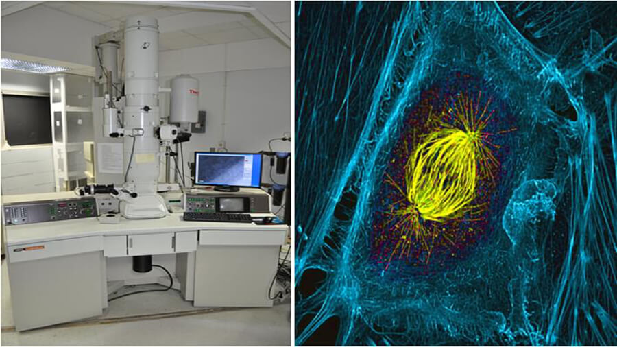
TEM is a microscopy technology technique where an electron beam is transmitted through ultrathin specimen, interacting with the specimen as the electron beam passes through it, resulting in the formation of living image. Then the image is magnified and focused onto an imaging device, such as a fluorescent screen, on a layer of photographic film, or to be detected by a sensor such as a CCD camera. All samples prepared for TEM must be small and thin enough to be electron transparent, which is comparable to the mean free path of the electrons that travel through the samples. In addition, all water must be removed from the samples as the TEM requires vacuum environment.
Some products (such as films and wax) have different sizes: please feel free to contact us for any inquiries. Creative Biostructure also provides various custom UniCrys™ membrane protein crystallization services and products for SEM and cryo-EM. Please feel free to contact us for a detailed information.
References:
M. A. Haque and M. T. A. Saif (2001). In-situ tensile testing of nano-scale
specimens in SEM and TEM.
Experimental Mechanics
,
42
: 123.
Transmission electron microscopy. (https://en.wikipedia.org/wiki/Transmission_electron_microscopy).


