DNA Structure Characterization
DNA is a polymer of nucleotides, consisting of a five-carbon sugar and phosphate backbone and nitrogenous bases, and forms a double helix structure. The double helix structure has various conformations such as A, B, and Z. DNA is the genetic material of living organisms and the conformational dynamics of DNA is increasingly recognized as a factor influencing its biomolecular function. Therefore, high-resolution techniques are essential to reveal fundamental biomolecular insights including the structural features of DNA, thereby facilitating understanding by researchers and enabling general applications.

- Nuclear magnetic resonance (NMR) spectroscopy
- Fluorescence resonance energy transfer (FRET)
- Single-molecule (sm) FRET
- Single-pair (sp) FRET
- Raman spectroscopy
- SERS
- Circular dichroism (CD) spectroscopy
NMR spectroscopy is a widely used technique because of its ability to provide atomic insight under near-physiological conditions. Solution-state NMR spectroscopy is a unique technique that maps both structural and conformational fluctuations in DNA molecules, playing an integral role in testing hypotheses and detecting subtle changes that are not easily detectable.
FRET as an optical molecular ruler in the 1.0-10.0 nm distance range can be performed in living cells with high resolution, high sensitivity, simplicity, and convenience. Its increasingly widespread use is in nucleic acid detection, immunoassays, and the study and structural characterization of biomolecular interactions, such as DNA.
X-ray crystallography, NMR spectroscopy, and cryo-electron microscopy (cryo-EM) are the gold standard for determining the structure of biomolecules. However, these techniques are often unable to observe small conformational changes in biomolecules as the required sample preparation may stabilize a particular conformation. In contrast, smFRET can be used to determine molecular structures at the sub-nanometre resolution, including rare conformations.
Intracellular spFRET allows quantitative in vivo characterization of DNA structure without significant interference with the natural cellular environment. Compared to bulk intracellular FRET measurements, quantitative spFRET measurements avoid the need to introduce large amounts of DNA into the cell and the use of chemical transfection agents, and will greatly diminish interference with the composition of the intracellular environment.
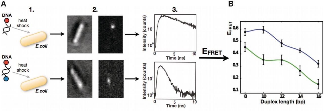 Figure 1. Simplified workflow of the in-cell spFRET experiment. (Tomáš, F., et al., 2012)
Raman spectroscopy is a non-destructive analytical technique based on the interaction of light and chemical bonds within a material. The technique has many advantages such as its non-destructive nature, short detection time, the small sample size required, and simple sample preparation. With the introduction of laser light sources, the improvement of weak detection techniques, and the use of computers, several new techniques have emerged, such as Fourier Transform Raman Spectroscopy (FT2Raman), Resonance Raman Spectroscopy (RRS), Surface Enhanced Raman Spectroscopy (SERS) and Confocal Micro Raman Spectroscopy (CRM).
SERS is a promising method for label-free DNA analysis, as it is highly sensitive and provides information on chemical structure.
CD spectroscopy is a powerful tool for characterizing the structure of DNA in solution. although it does not provide high-resolution structural information, it is very sensitive to DNA secondary structure and is particularly suitable for monitoring structural changes caused by folding, binding and environmental perturbations. the secondary structure of DNA (type A, B, Z, etc.) has a characteristic CD spectrum.
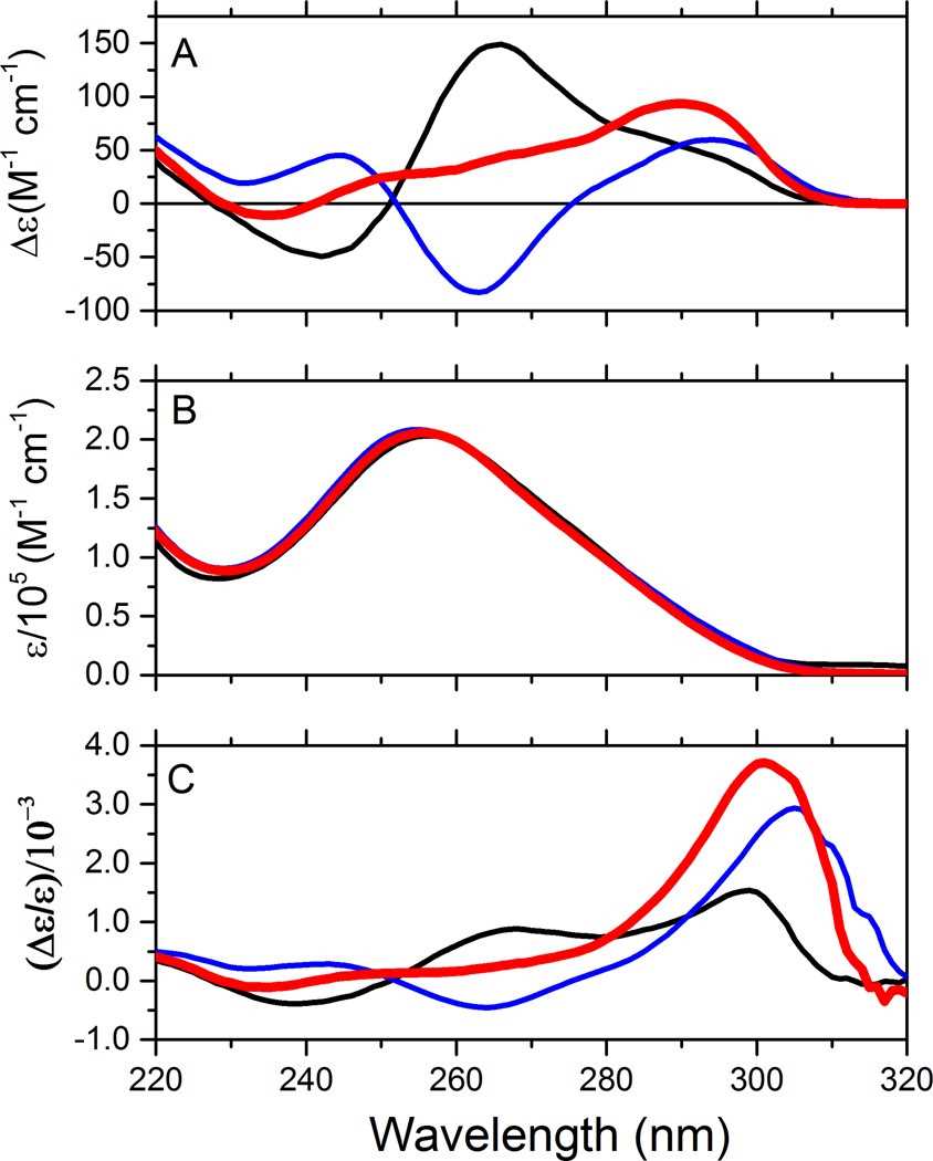 Figure 2. Spectral features of human telomer quadruplex conformations. (Del Villar-Guerra, R., et al., 2017)
- DNA Origami and Self-Assembly Service
DNA origami technology is an innovative nanotechnology that leverages the inherent properties of DNA molecules to create nanostructures with specific shapes and functions. Creative Biostructure has consistently focused on the analysis of biological structures, offering state-of-the-art DNA origami technology and self-assembly services.
Please feel free to contact Creative Biostructure for more details on our DNA structure characterization projects.
Ordering Process

References
- Fessl T., et al. Towards characterization of DNA structure under physiological conditions in vivo at the single-molecule level using single-pair FRET. Nucleic Acids Research. 2012, 40(16): e121-e121.
- del Villar-Guerra R., et al. Characterization of quadruplex DNA structure by circular dichroism. Current Protocols in Nucleic Acid Chemistry. 2017, 68(1): 17.8. 1-17.8. 16.


