Inquiry Basket
| Your inquiry basket is currently empty. |
Combine morphology, particle size and concentration, marker identity, and optional molecular analysis in a project-specific characterization plan.
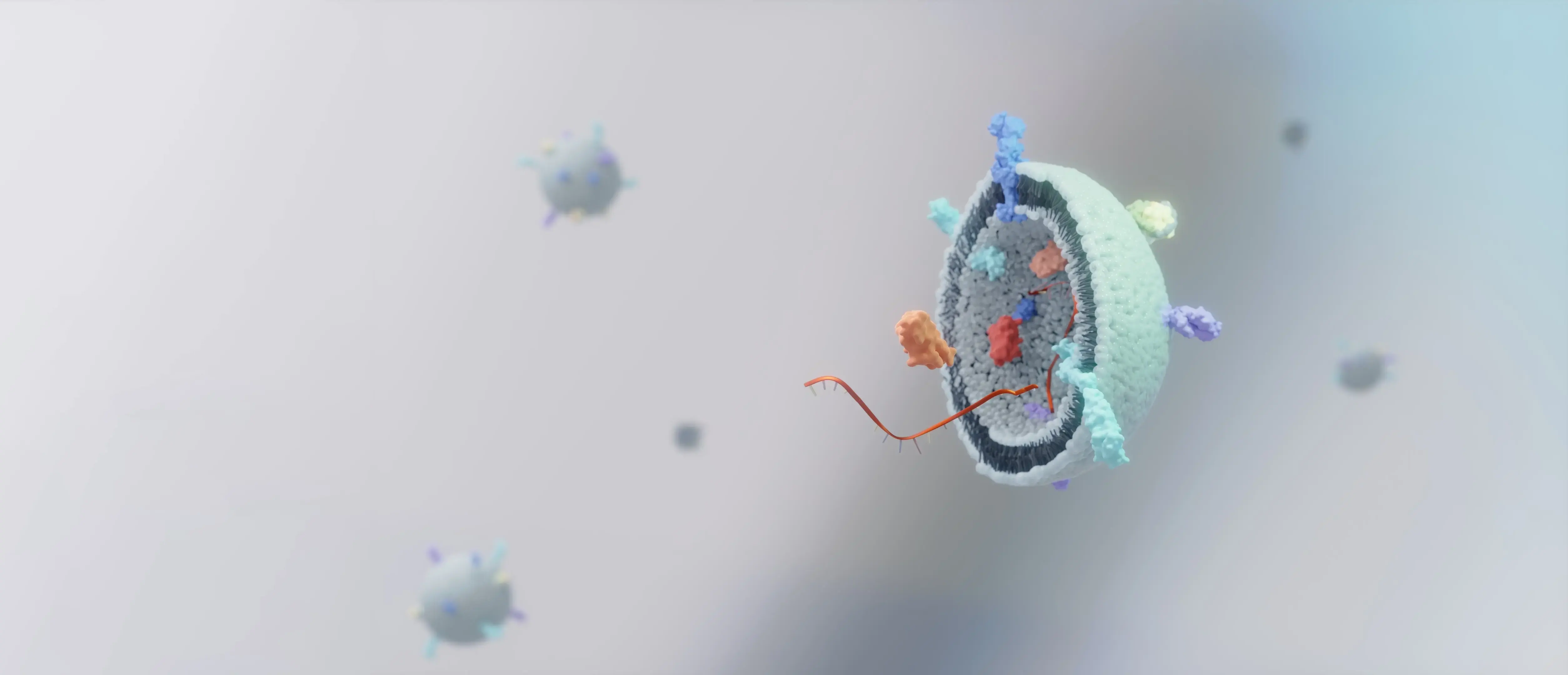
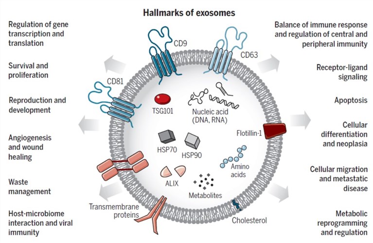
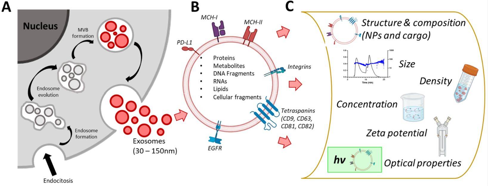
Exosome characterization combines complementary analytical methods to assess vesicle morphology, size distribution, particle concentration, and marker identity. It matters because no single measurement can establish exosome identity or determine whether a preparation is suitable for downstream research.
Exosomes are heterogeneous nanoscale vesicles. A robust characterization plan should examine complementary properties instead of relying on a single readout.
Creative Biostructure typically organizes characterization at three levels: morphology, size and size distribution, and surface-marker identification. Optional methods can extend the analysis to particle concentration, surface properties, cargo, or molecular composition.
Morphology shows particle appearance; NTA, DLS, or TRPS describe particle behavior and size; marker assays evaluate proteins associated with the vesicle preparation.
Each technique answers a different question and has distinct limitations. Combining readouts reduces overinterpretation and creates a more defensible description of the sample.
Results can inform preparation comparison, quality review, method selection, downstream assay planning, and reporting for exosome research projects.
Characterization capabilities
Begin with the property you need to establish, then combine complementary methods to strengthen interpretation.
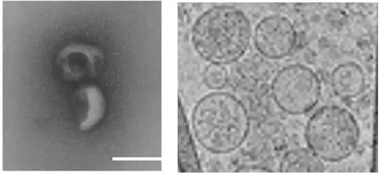
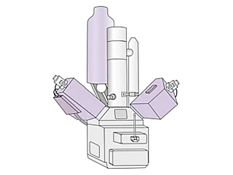
Visualize vesicle morphology, membrane integrity, and particle appearance.
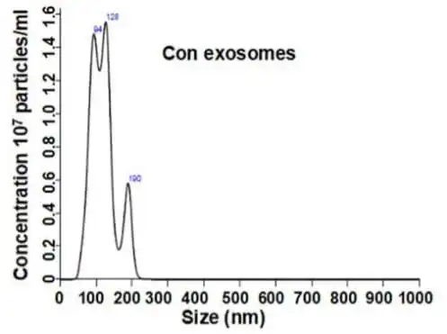
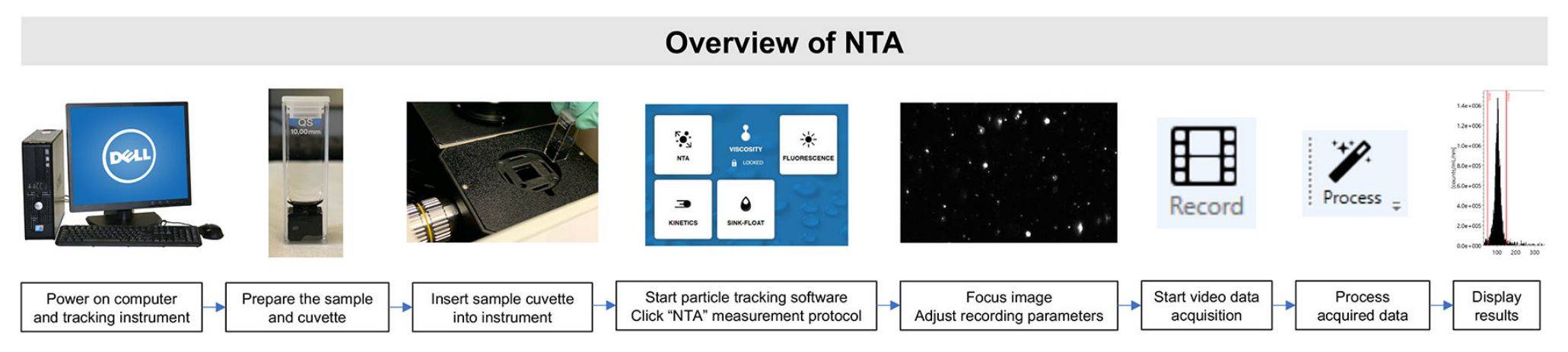
Measure particle size distribution and concentration in liquid suspension.
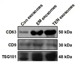
Evaluate CD9, CD63, CD81, TSG101, ALIX, HSP70, and project-specific markers.
Add complementary size, surface, biomechanical, or molecular-composition data.
Method selection guide
No single assay answers every characterization question. Use the comparison below to identify an appropriate starting panel.
| Research question | Recommended method | Primary readout | Important consideration |
|---|---|---|---|
| What do the vesicles look like? | TEM / Cryo-TEM / SEM | Morphology and structural appearance | Preparation influences interpretation |
| What is the size distribution and concentration? | NTA | Particle size profile and count | Does not prove exosome identity |
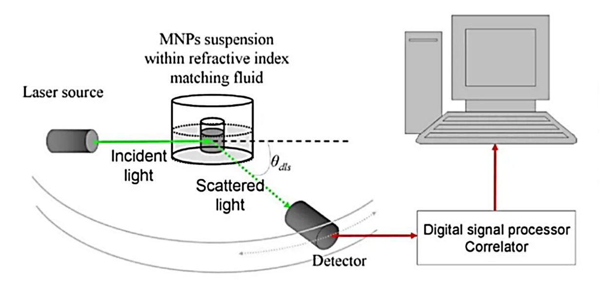
| Is the suspension relatively monodisperse? | DLS | Hydrodynamic size and distribution | Best for relatively uniform suspensions |
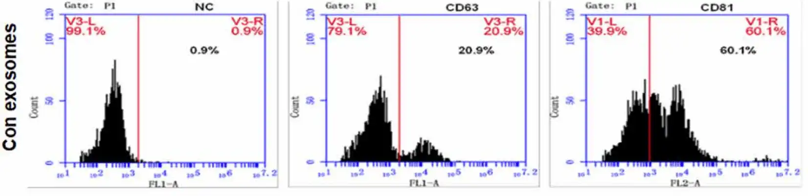
| Which vesicle markers are present? | Western blot / Flow Cytometry / FACS | Marker detection or expression profile | Panel should fit the sample source |
| What is the molecular composition? | Mass spectrometry | Protein or molecular-composition data | Scope depends on sample and objective |
Send your sample source, isolation method, available amount, and downstream objective.
| Capability | Details |
|---|---|
| Morphology
& Ultrastructure |
TEM, Cryo-TEM, and SEM visualize vesicle morphology, membrane preservation, particle appearance, and aggregation. Imaging strategy and sample preparation are selected to fit the question. |
| Particle Size
& Concentration |
NTA measures particle-size distribution and concentration. DLS and TRPS can add complementary hydrodynamic-size or single-particle information when appropriate. |
| Marker Identity
& Phenotyping |
Western blot, flow cytometry/FACS, and immunoelectron microscopy evaluate exosome-associated and project-specific markers using a panel matched to the sample source. |
| Surface & Nanoscale Properties |
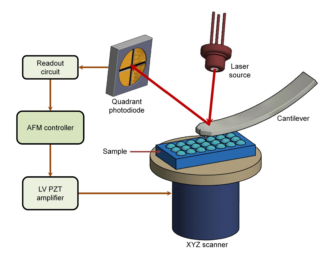
AFM provides complementary surface-topography and nanoscale-property information for projects that require more than routine morphology and particle analysis. |
| Molecular Composition | Mass spectrometry can extend the study to protein or broader molecular-composition questions, with analytical scope defined by sample quality, available material, and the research objective. |
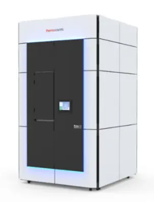
Thermo Scientific Krios G4
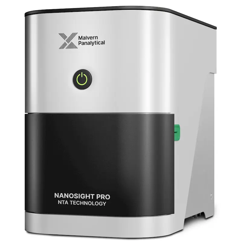
NanoSight Pro NTA particle size analyzers
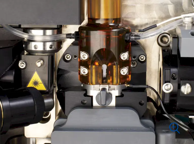
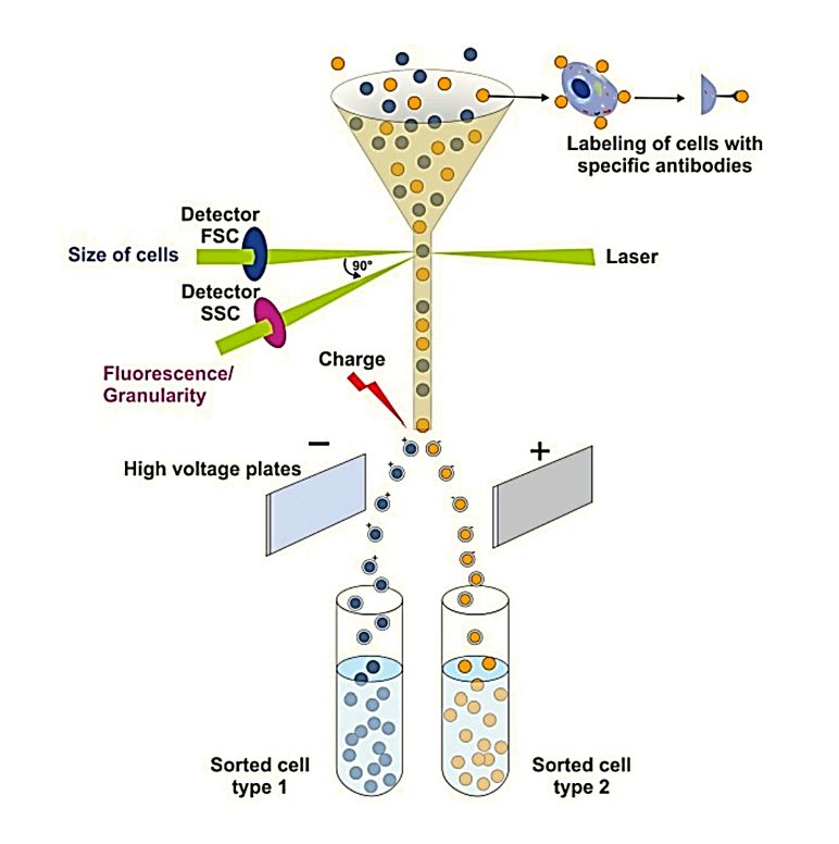
BD FACSAria™ Fusion Flow Cytometer
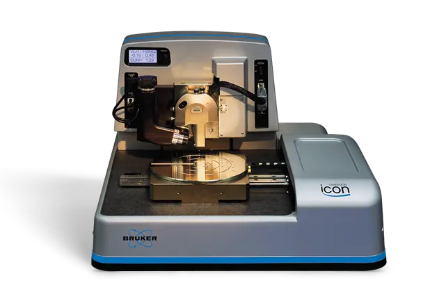
Bruker Dimension Icon AFM
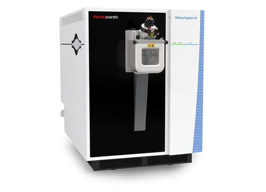
Thermo Scientific Orbitrap Exploris 480
Project workflow
Each method is selected to answer an explicit scientific question, with limitations and outputs defined before testing begins.
Sample source, preparation history, prior results, and downstream goal.
Confirm method fit and project-specific handling requirements.
Perform agreed morphology, particle, marker, or composition methods.
Review controls, consistency, and assay-specific limitations.
Organize results around the agreed research question.
Clear project outputs
Deliverables are defined with the selected methods and may include representative images, plots, marker data, method summaries, and scientific interpretation.
Method-specific resultsImages, profiles, plots, blots, or datasets according to the agreed panel.
Quality and interpretation notesContext for controls, assay limitations, and complementary evidence.
Organized project reportA consolidated record of methods, results, and project-specific conclusions.
Client publication case study
A peer-reviewed study used transmission electron microscopy performed at Creative Biostructure to check extracted hucMSC exosomes before evaluating their effects in an in vitro cardiomyocyte injury model.
Hypoxia/reoxygenation can activate the NLRP3 inflammasome and drive inflammatory cell death in cardiomyocytes. The investigators asked whether human umbilical cord mesenchymal stem cell-derived exosomes could reduce this injury and whether exosomal miR-100-5p acted through the FOXO3/NLRP3 pathway.
Frequently asked questions
Requirements depend on the methods selected. A short technical review is the fastest way to define the right panel.
Creative Biostructure offers NTA, TEM and Cryo-TEM, SEM, Western blot, flow cytometry/FACS, DLS, TRPS, AFM, immunoelectron microscopy, and mass spectrometry. The recommended panel depends on the sample and research objective.
Publications
These publications provide traceable scientific context for exosome characterization and document Creative Biostructure services or contributions cited by researchers.
Al-shubaily, F. A., & Al-Zahrani, M. H. (2021). In Role of Exosomes in Biological Communication Systems, 27-75.
Scientific chapter discussing exosome structure and characterization approaches, with reference to Creative Biostructure characterization technology.
Liang, C., et al. (2021). Frontiers in Bioengineering and Biotechnology, 8, 615850.
The authors report that extracted hucMSC exosomes were checked by TEM at Creative Biostructure before functional testing.
Bickmore, D. C., & Miklavcic, J. J. (2020). Frontiers in Nutrition, 7, 22.
The publication reports fatty acid analysis performed by Creative Biostructure as part of a human-milk extracellular-vesicle study.
Related services
Research resources
Move from service evaluation to practical planning with downloadable brochures, research examples, and technical support.

References










For Research or Industrial Raw Materials, Not for Personal Use!
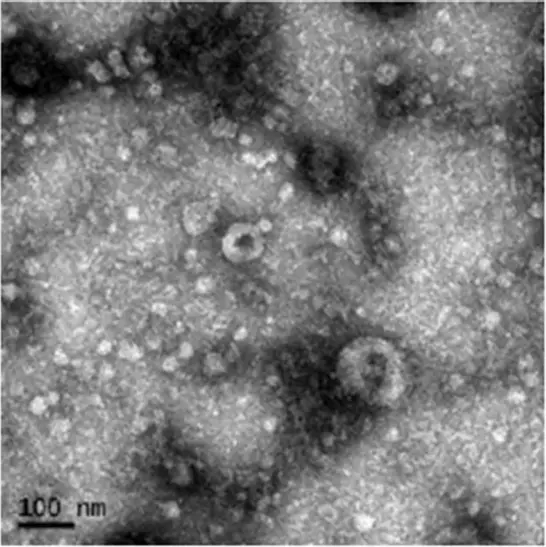 Figure 1. Morphology of purified hucMSC-exo. Scale bar: 100 nm