Particle Morphology Analysis
Cryogenic electron microscopy (cryo-EM) particle analysis technology has become a new force in structural biology, and it is playing an increasingly important role in the structural analysis of mixed liposome and exosome nanoparticles. Basic information about the nature and source of particulate matter can usually be gleaned from the size, shape, and/or surface form of individual particles.
Employing cryo-EM, we can analyze the particle morphology and obtain the protein structures when they bind to small molecules, which plays an important role in the development of pharmaceutical chemistry. We are working hard to study with the world's top laboratories and build a cryo-EM team. Our particle morphology analysis is based on the following principles: When a frozen sample of a tiny particle is placed under the EM, it is subjected to non-destructive bombardment by an accurately focused electron beam (probe). The most commonly used emission signals in particle morphology analysis are secondary electrons, characteristic X-rays and backscattered electrons. Secondary electrons generated from frozen particle samples are often used to characterize particle morphology. When a sample is irradiated by a primary electron stream, a variety of interactions occur with atoms in the sample. As a result, the sample emits various forms of radiation, which can be used to determine its composition after testing and processing.
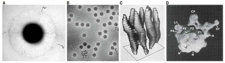 Figure 1. Establishment of the single particle cryo-EM.
Our EM platform develops a particle morphology characterization system that can analyze the shape and size of individual particles with particle sizes ranging from 40 nm to 2,000 nm, distinguish individual particles or polymers, quantify structures in polymers, analyze spheres or non-spheres, and count individual particles in mixtures. We can conduct particle morphology analysis on the nanoparticles, calculate multiple particle shape parameters for each particle, and generate distribution according to each parameter. The equivalent diameter, length, width, perimeter, area, aspect ratio, roundness, protrusion, and real product of the circle can be given, where the aspect ratio refers to length divided by width, the protrusion refers to the circumference of the convex enclosure divided by the actual circumference, and the real product refers to the actual area divided by the area of the convex enclosure.
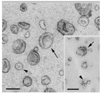 Figure 2. Exosome characterization using cryo-EM. (Théry C, et al. 2006)
Our cryo-EM team has rich experience in the processing and identification of lipid nanoparticles, liposomes, exosomes, emulsion, and virus-like particles.
Our service advantages:
- We have many years of experience in cryo-EM technology.
- Our samples are handled with fully automated, highly repeatable equipment and precise control of key process parameters.
- We can conduct high throughput sample testing service.
- Simple and fast user interface, timely after-sales service
If you are interested in our particle morphology analysis service, please feel free to contact us. We are looking forward to cooperating with you.
Ordering Process

Reference
- Théry C, Amigorena S, Raposo G, et al. Isolation and characterization of exosomes from cell culture supernatants and biological fluids. Current Protocols in Cell Biology. 2006, 30(1): 3.22. 1-3.22. 29.


