Inquiry Basket
| Your inquiry basket is currently empty. |
From AI-predicted hypotheses to experimentally validated molecular structures.
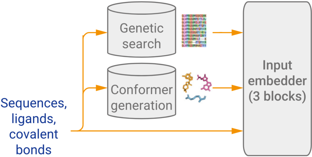
Creative Biostructure integrates sequence analysis, construct design, protein production, X-ray crystallography, cryo-EM, NMR, EPR spectroscopy, biophysical assays and computational refinement in one iterative workflow.
Structure prediction, construct mapping, docking and dynamics prioritize testable questions.
Sample state, method, controls and acquisition strategy are selected around the question.
X-ray, cryo-EM, NMR, MicroED and EPR reveal structure, distance restraints and dynamics.
Experimental constraints update the model and direct the next construct or experiment.
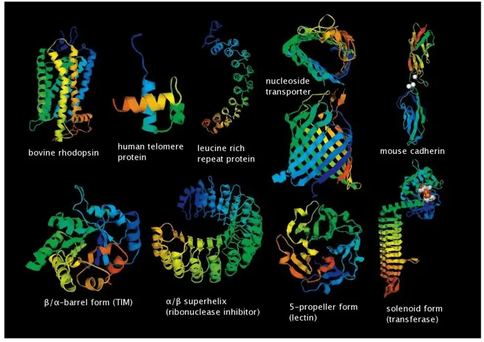
It connects molecular form with biological function. Structural evidence can reveal binding pockets, interfaces, catalytic arrangements, oligomeric states, flexibility and conformational change that are not fully resolved from sequence alone.
No single method answers every structural question. X-ray crystallography can provide atomic detail for crystallizable samples; cryo-EM is powerful for large or heterogeneous assemblies; NMR probes solution-state structure and dynamics; microscopy and biophysical assays add spatial, kinetic and thermodynamic evidence. Integrative structural biology combines these data types around one biological question.
Where are the atoms, domains or subunits?
How do partners, ligands or antibodies bind?
Which states change with function or environment?
Computational models narrow the experimental search space. Wet-lab measurements test those hypotheses. The resulting evidence then guides the next design decision.
Translate the biological objective into a resolvable question: architecture, binding mode, interface, dynamics, state or mechanism.
Use domain boundaries, disorder, transmembrane regions, conservation and predicted structure to design testable constructs.
Express, purify and assess identity, purity, monodispersity, stability, activity and complex formation before acquisition.
Predicted models, docking and simulations help rank constructs, ligands, conformations and conditions—not replace physical testing.
Combine coordinate or density methods with binding, stability and EPR distance-distribution data where dynamics or heterogeneity matter.
Fit experimental constraints, review validation metrics, reconcile disagreements and feed insights into the next design cycle.
AI predictions are probabilistic. Experimental structures are also method- and sample-dependent. Iteration makes assumptions visible and improves confidence through cross-validation.
Choose a standalone method or combine complementary technologies to resolve structure, interaction and dynamics across scales.
Atomic-level structures of crystallizable proteins, complexes and protein–ligand systems, from screening through model refinement.
Single-particle and electron microscopy workflows for large complexes, membrane proteins and conformational heterogeneity.
Solution- and solid-state analysis of structure, binding, flexibility and molecular dynamics across relevant timescales.
Diffraction-based structure determination for microcrystalline proteins, peptides and small molecules.
Site-specific distance distributions, conformational populations and protein dynamics using spin-label measurements.
Joint interpretation of cryo-EM, X-ray, NMR, mass spectrometry, microscopy and modeling constraints.
Spatial organization, localization and dynamic behavior in cells at resolution beyond conventional light microscopy.
Structure prediction, model reconstruction, docking, dynamics, data processing and evidence-based interpretation.
The best method depends on the decision, not only molecular weight. Sample behavior, flexibility, environment, resolution needs and available material all influence strategy.
| Method | Strong fit | Information gained | Important constraint | Typical output |
|---|---|---|---|---|
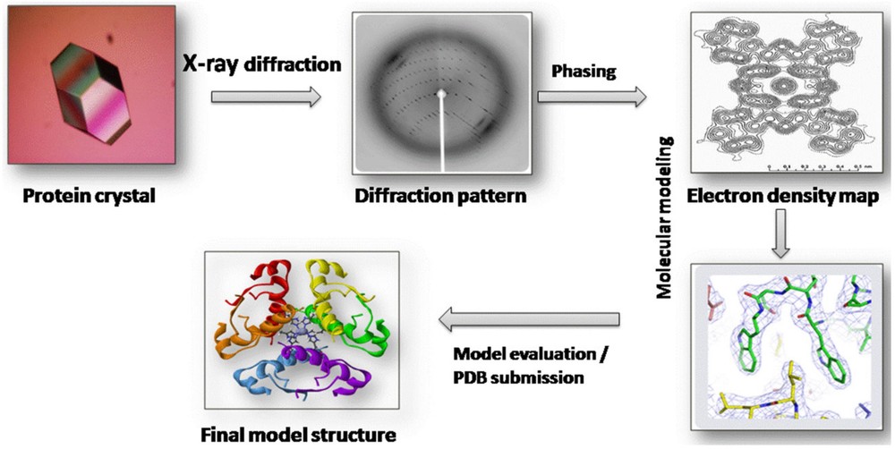
| X-ray crystallography | Well-ordered soluble proteins and complexes | High-resolution atomic coordinates and ligand interactions | Requires crystals; conformational ensembles may be underrepresented | Diffraction data, refined coordinates, validation statistics |
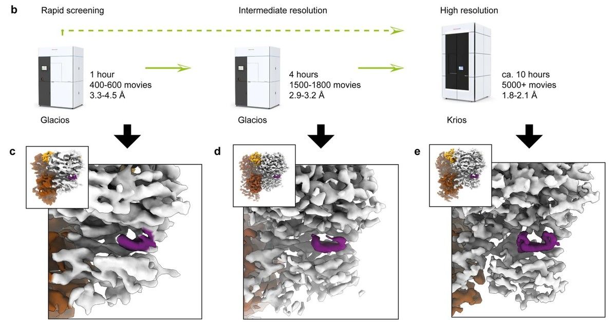
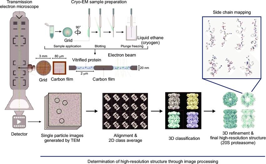
| Cryo-EM / SPA | Large assemblies, membrane proteins, multiple states | 3D density, architecture and conformational classes | Grid behavior, preferred orientation and heterogeneity affect resolution | Micrographs, 2D classes, density maps, atomic model where supported |
| NMR spectroscopy | Smaller proteins, flexible regions, binding and dynamics | Solution-state structure, chemical environment and motion | Size, labeling, concentration and spectral overlap can limit analysis | Spectra, assignments, restraints, structures and dynamics metrics |
| MicroED | Microcrystalline peptides, proteins and small molecules | Diffraction-based 3D structure from very small crystals | Crystal thickness, radiation damage and data completeness matter | Diffraction data and refined structural model |
| EPR spectroscopy | Flexible proteins, membrane proteins, complexes and disordered systems with suitable spin-label sites | Nanometer-scale distance distributions, conformational populations and changes in dynamics | Requires strategic labeling and does not by itself produce a complete atomic structure | Raw traces, distance-distribution profiles, restraints and model comparison |
| Integrative approach | Flexible, heterogeneous or multi-component systems | Cross-scale model constrained by complementary evidence | Requires explicit uncertainty and compatibility assessment across datasets | Evidence-weighted ensemble or architecture with provenance |
Binding, stability, size and conformational assays—including EPR distance distributions—help test whether a structural model represents the relevant molecular state or ensemble.
SPR, BLI, ITC, MST and fluorescence-based assays can confirm affinity, kinetics, stoichiometry and thermodynamic behavior.
DSC, thermal shift, DLS, MALS, SEC and AUC can assess folding, aggregation, molecular weight and oligomeric state.
HDX-MS, EPR, CD, FTIR and complementary spectroscopy can map flexibility, secondary structure and environment-dependent changes.
Site-directed spin labeling and pulsed dipolar EPR can provide probability distributions of inter-spin distances. These restraints can test predicted conformations, refine low-resolution maps and compare ligand- or environment-dependent states.
Projects may begin with a sequence, construct, purified sample, complex or existing dataset.
Soluble and membrane proteins, receptors, enzymes, antibodies, fragments, oligomers and disordered regions.
Protein–protein, protein–ligand, protein–peptide, antibody–antigen and macromolecular assemblies.
Active/inactive states, folding intermediates, transient interactions, allosteric transitions and assembly pathways.
Automated preparation, high-resolution acquisition and expert data analysis support projects from screening through validated models.
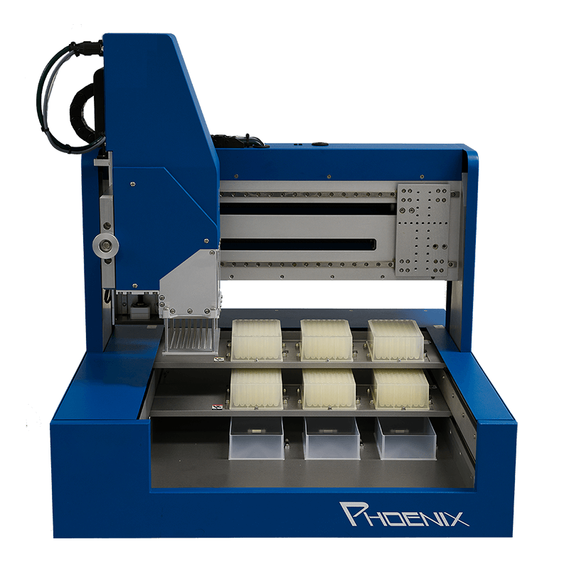
High-throughput screening and nanoliter dispensing.
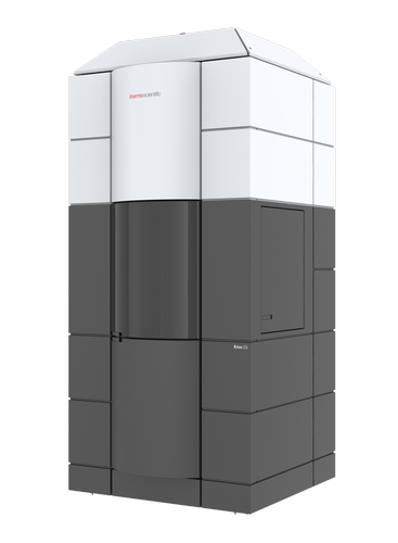
High-resolution imaging of complexes and membrane proteins.
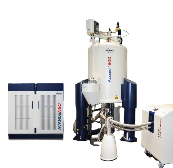
Solution and solid-state structural measurements.
Processing pipelines with transparent validation metrics.
Exact purity, concentration, quantity, buffer and activity requirements vary by target and method.
Review experimental images, structural outputs and downloadable summaries from representative X-ray crystallography and Cryo-TEM projects.
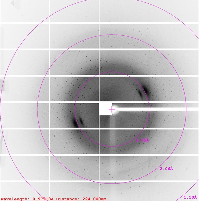
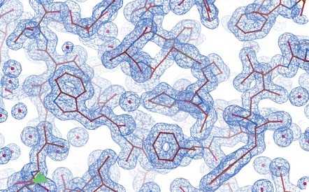
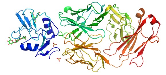
This project focuses on elucidating the three-dimensional structure of a Fab–antigen complex using X-ray crystallography. The workflow begins with Fab preparation, complex formation and initial crystal screening, followed by diffraction analysis, density-map interpretation and model refinement.
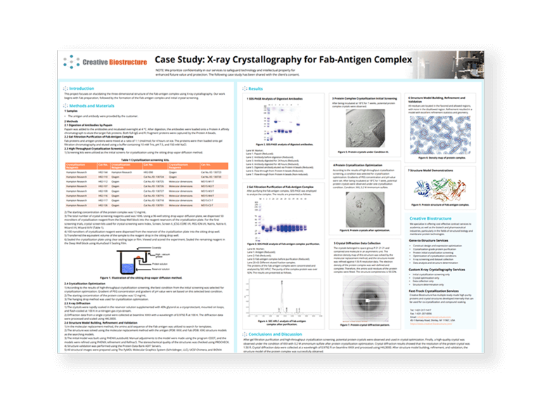
Complete the short form to access the full background, methods, results and structural interpretation.
This project analyzes the three-dimensional structure of a CDXX receptor and AB-X antibody complex using Cryo-TEM. Sample preparation is followed by data acquisition, image processing, single-particle classification, model building and refinement.
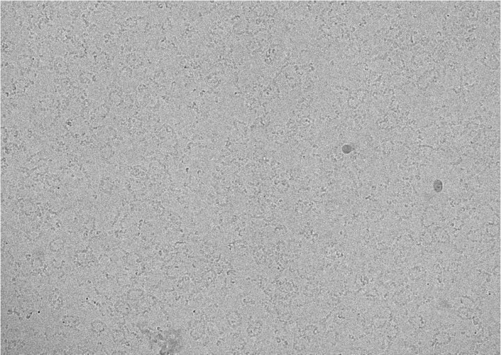
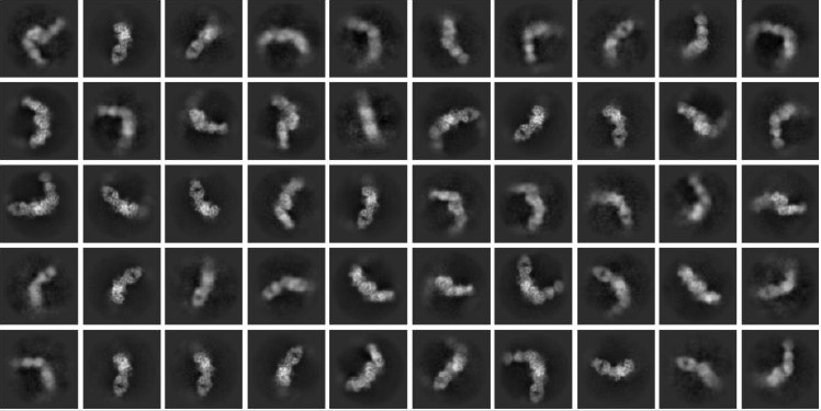
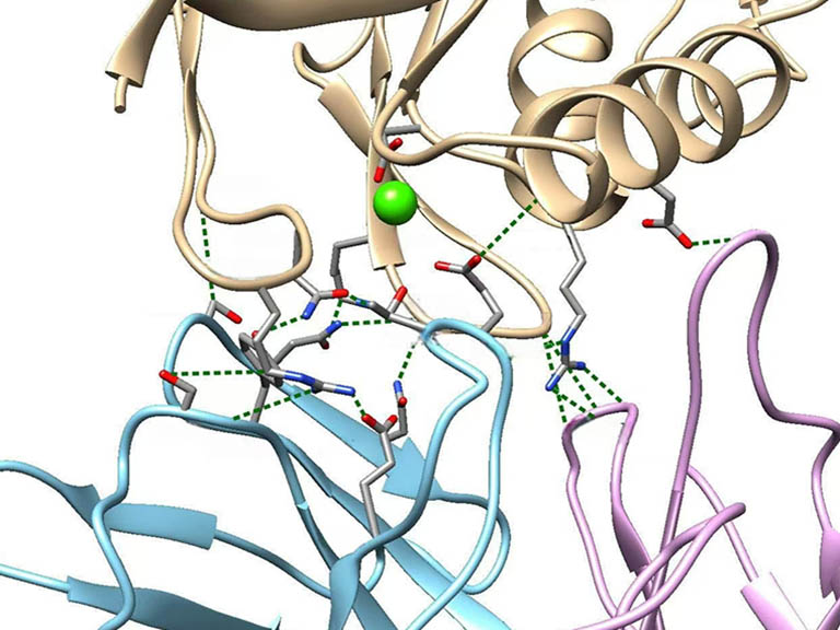
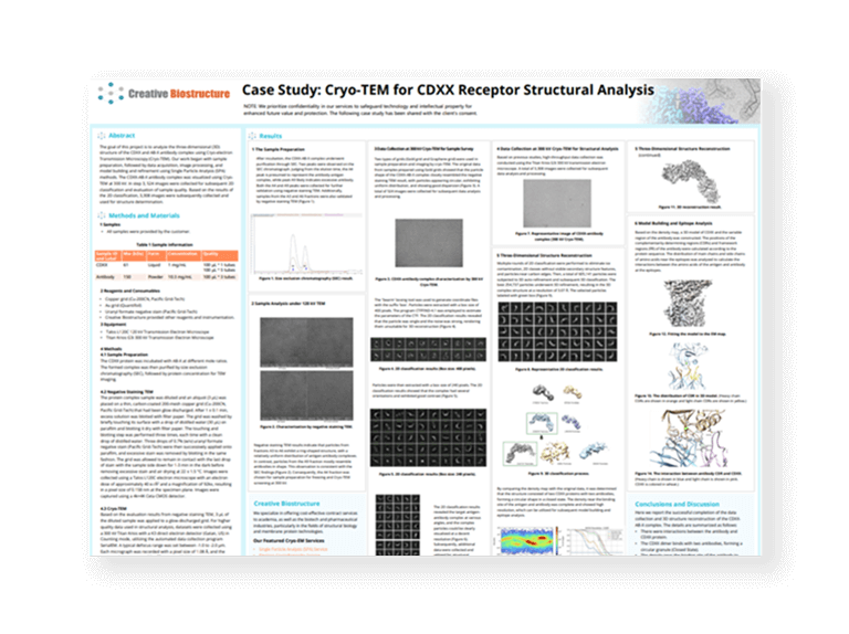
Complete the short form to access the workflow, representative images and structural insights.
Structural evidence supports target assessment, molecular engineering and mechanistic interpretation across discovery and development programs.
Explore how structural determination, computational modeling and interaction analysis can support target assessment and drug-design decisions.

Review our integrated capabilities for protein and macromolecular structure analysis, illustrated through a membrane-protein project example.
The studies below identify specific structural biology contributions from Creative Biostructure. Article titles, journals, years and contribution summaries are presented together for direct review.
| Article | Journal | Year | Contribution |
|---|---|---|---|
| APOE Christchurch-mimetic therapeutic antibody reduces APOE-mediated toxicity and tau phosphorylation | Alzheimer's & Dementia | 2024 | Creative Biostructure determined the three-dimensional crystal structure of purified 7C11.IgG Fab as a fee-for-service contribution. |
| A novel mechanism of herbicide action through disruption of pyrimidine biosynthesis | Proceedings of the National Academy of Sciences | 2023 | Creative Biostructure provided X-ray crystallographic support for the structural investigation reported in the study. |
| Linoleic acid binds to SARS-CoV-2 RdRp and represses replication of seasonal human coronavirus OC43 | Scientific Reports | 2022 | The work used a Creative Biostructure-produced complex structure of SARS-CoV-2 RNA polymerase with double-stranded RNA fragments. |
| Synthesis and biological evaluation of selective survivin inhibitors derived from the MX-106 hydroxyquinoline scaffold | European Journal of Medicinal Chemistry | 2021 | The authors report collaboration with Creative Biostructure to optimize survivin crystallization conditions for inhibitor-complex structure determination. |
| All major cholesterol-dependent cytolysins use glycans as cellular receptors | Science Advances | 2020 | The authors acknowledge Creative Biostructure for protein chemical-shift assignment supporting the study. |
Clear answers to common questions about prediction, method choice, samples and deliverables.
View all FAQs →No. AI models are valuable hypotheses, but experiments are needed to verify the physical sample state, ligand pose, local geometry, dynamics, oligomeric state and method-specific uncertainty.
Method selection depends on molecular size, sample homogeneity, flexibility, available quantity, crystallization behavior, environment and the structural or dynamic information required. Some questions benefit from an integrative strategy.
A project may combine detergent or lipid screening, stability and monodispersity assessment, nanodisc or liposome reconstitution, and cryo-EM, X-ray or NMR analysis selected for the target state.
EPR uses site-specific paramagnetic labels to provide nanoscale distance distributions and conformational-population information. These restraints can test predicted models, complement low-resolution density and reveal flexible or multiple states that a single static structure may not capture.
Projects can begin from a gene sequence, construct, expression system, purified protein, nucleic acid, complex or an existing structural dataset. Exact specifications are assessed for the target and method.
Depending on scope, deliverables may include quality-control results, raw and processed data, density maps or spectra, refined coordinates, validation metrics, interaction analysis, figures and a technical report.
Yes. Existing predictions can be treated as testable starting models and evaluated against structural, binding, stability or dynamics data. The validation plan should define which claims the experiments can and cannot support.











For Research or Industrial Raw Materials, Not for Personal Use!