The adaptive immune system protects the body by identifying and eliminating infected or abnormal cells. Central to this process are T lymphocytes (T cells), which recognize specific antigen fragments known as T cell epitopes through their T cell receptors (TCRs). Decoding how T cells recognize these epitopes is essential for advancing immunotherapies, vaccines, and immune-related research.
T cell epitopes are short peptides presented by major histocompatibility complex (MHC) molecules for TCR recognition. Accurate prediction and experimental mapping of T cell epitopes are critical for vaccine design, cancer immunotherapy, and autoimmune disease research. This article outlines the fundamental concepts of T cell epitopes, key methods for their prediction and mapping, and their applications in biomedical science.
What is a T Cell Epitope
A T cell epitope is a short linear peptide fragment derived from an antigen, such as a viral, bacterial, or tumor-associated protein, that is specifically recognized by a T cell receptor (TCR). Unlike B cell epitopes, which are often three-dimensional surface structures recognized by antibodies, T cell epitopes are typically linear sequences presented by major histocompatibility complex (MHC) molecules for T cell activation.
For a deeper understanding of how B cell epitopes are identified and analyzed, refer to Overview of B Cell Epitope Prediction and Mapping.
How MHC Molecules Present Antigenic Epitopes
T cells do not directly recognize intact antigen molecules. Instead, they recognize antigenic peptide fragments presented by MHC molecules on the cell surface. MHC molecules are broadly classified into two types:
| MHC Class I Molecules | MHC Class II Molecules |
|---|---|
| Found on the surface of nearly all nucleated cells. They primarily present short peptides (typically 8-11 amino acids) derived from intracellular endogenous antigens (e.g., viral proteins, tumor proteins) that have been degraded by the proteasome and transported into the endoplasmic reticulum via the TAP transporter. | Primarily found on professional Antigen-Presenting Cells (APCs), such as dendritic cells, macrophages, and B cells. They mainly present longer peptides (typically 13-17 amino acids, sometimes up to 25 or more) derived from exogenous antigens (e.g., bacteria, parasites) that have been taken up by endocytosis and processed in lysosomes. |
Antigen processing and MHC presentation are prerequisites for T cell epitopes to exert their function.
 Figure 1. CD4⁺ T-Cell Recognition of Antigen Peptides Presented by MHC II. (Sanchez-Trincado J L, et al., 2017)
Types of T Cell Epitopes
Based on which T cell subset recognizes them and the class of MHC molecule presenting them, T cell epitopes are mainly categorized as:
| CD4+ T Cell Epitopes | CD8+ T Cell Epitopes |
|---|---|
| Presented by MHC Class II molecules and recognized by helper T lymphocytes (Th cells). Th cells regulate the functions of other immune cells (e.g., B cells producing antibodies, CTL activation, macrophage activation) primarily through cytokine secretion. | Presented by MHC Class I molecules and recognized by cytotoxic T lymphocytes (CTLs). CTLs can directly kill target cells (like virus-infected cells or tumor cells) expressing the corresponding epitope. |
 Figure 2. CD8⁺ T-Cell Recognition of Antigen Peptides Presented by MHC I. (Sanchez-Trincado J L, et al., 2017)
Key T Cell Epitope Properties
Whether a peptide segment can serve as an effective T cell epitope depends on several factors:
- Amino Acid Sequence: Sequence features influence its susceptibility to proteasomal cleavage, its ability to bind complementarily to anchor residues within the MHC binding groove.
- MHC Binding Affinity: The strength of binding (often measured by Kd or IC50 values) between the peptide and a specific MHC allele is a necessary condition for becoming an epitope. Higher affinity generally implies more effective presentation.
- Peptide-MHC Complex Stability: The formed complex must remain stable on the cell surface long enough for effective TCR recognition.
- TCR Recognition: Even if a peptide binds MHC, its exposed residues must be recognizable and bindable by an appropriate TCR.
- Immunogenicity: Refers to the actual ability of a T cell epitope to induce a detectable T cell response, such as proliferation, cytokine secretion, or cytotoxicity. This represents the ultimate measure of an epitope's functional relevance.
- Conservation: For pathogen or tumor antigens, the conservation of an epitope sequence across different strains or tumor samples is critical for its potential as a broadly applicable vaccine or therapeutic target.
Why Predict and Map T Cell Epitopes
- Fundamental Immunology Research: Deepening the understanding of T cell recognition mechanisms, immune response diversity, and regulation.
- Epitope-based Vaccine Development: Designing epitope-based vaccines that precisely target immune responses to critical, conserved regions of pathogens or tumors, enhancing vaccine efficacy and safety.
- Immunotherapy: Developing personalized cancer vaccines or adoptive T cell therapies (ACT) targeting tumor-specific neoantigens; identifying pathogenic epitopes in autoimmune diseases for developing tolerance-inducing therapies.
- Infectious Disease Research: Understanding the host T cell immune response characteristics to specific pathogens, tracking the impact of viral mutations on immune recognition.
- Biotherapeutic Safety Assessment: Predicting and assessing the potential immunogenicity risk of protein-based drugs (e.g., antibody drugs, enzyme replacement therapies), guiding drug optimization (deimmunization).
T Cell Epitope Prediction: In Silico Identification Strategies
Faced with the vast sequence information of proteins, T cell epitope prediction aims to use computational biology and bioinformatics methods to screen large numbers of candidate peptides and identify those most likely to bind specific MHC molecules and induce T cell responses. This in silico approach vastly reduces the number of antigenic peptides needing experimental testing, significantly saving time and cost, and providing strong guidance for subsequent experiments.
Core T Cell Epitope Prediction Approaches
MHC Binding Prediction
This is the cornerstone and most common method for T cell epitope prediction, as peptide binding to MHC is the first requirement for TCR recognition. Algorithms predict the binding affinity of peptides to specific MHC alleles. Common methods include:
- Sequence-based methods: Data-driven approaches like Position-Specific Scoring Matrices (PSSMs), Artificial Neural Networks (ANNs), and Support Vector Machines (SVMs), trained on large datasets of known binding/non-binding peptides (often sourced from databases like IEDB, EPIMHC, Antijen).
- Structure-based methods: Utilizing crystal structure information of MHC molecules and peptides to simulate the binding process through molecular docking, etc.
- Challenges and Differences: The high degree of polymorphism in human MHC (HLA) genes is a major prediction challenge. Furthermore, because the peptide-binding groove of MHC Class II molecules is open at both ends, accommodating peptides of variable length (13-25aa) and requiring accurate prediction of the 9-mer core binding region, prediction accuracy for Class II binding is generally lower than for MHC Class I molecules (which bind shorter 8-10aa peptides in a more closed groove).
Antigen Processing Prediction
Predicting whether a peptide can be efficiently processed within the cell. For MHC Class I epitopes, this involves predicting proteasomal cleavage sites and TAP transport efficiency; for MHC Class II epitopes, relevant protease cleavage sites are predicted.
Immunogenicity Prediction
This is a more challenging step. It attempts to predict the likelihood that an MHC-bound peptide will actually trigger a T cell response, potentially integrating information on peptide-MHC complex structural features, potential TCR interactions, the peptide's location within the antigen, etc.
Essential T Cell Epitope Prediction Tools and Databases
Numerous publicly available or commercial t cell epitope prediction tools and resources exist:
- IEDB (Immune Epitope Database and Analysis Resource): A comprehensive immune epitope database that also provides integrated prediction tools (Analysis Resource), including widely used algorithms like NetMHCpan (MHC Class I), NetMHCIIpan (MHC Class II), and MHCflurry. Its TepiTool workflow can integrate multiple prediction steps.
- Other Standalone Tools/Servers: Such as SYFPEITHI, BIMAS, RankPep, etc.
- Application of Artificial Intelligence: Machine learning models, particularly deep learning, are considered superior in silico prediction methods due to their ability to handle complex patterns and high-dimensional data, showing strong performance in peptide-MHC binding prediction.
Strengths and Limitations of Prediction
Strengths: Fast, low-cost, high-throughput, enabling rapid screening of entire proteomes. Prediction accuracy is relatively higher for T cell epitopes, which are mostly linear, compared to B cell epitopes that often involve discontinuous segments.
Limitations:
- Prediction accuracy is still limited, especially for immunogenicity, with both false positives and false negatives occurring.
- A gap exists between in silico predictions and the complex in vivo environment (e.g., actual antigen expression levels, processing efficiency, individual variations in T cell repertoire).
- Predictive capability for post-translationally modified peptides or non-classical epitopes is weaker.
- The extreme polymorphism of MHC alleles makes covering the entire human population challenging.
T Cell Epitope Mapping: Experimental Confirmation and Discovery
Due to the limitations of prediction, T cell epitope mapping—the experimental identification and validation of epitopes capable of inducing T cell responses—is an indispensable step. Its purpose is to confirm predictions, discover novel epitopes missed by algorithms, directly assess epitope function in actual biological samples (e.g., patient blood, vaccinee samples), and identify immunodominant epitopes. Experimental methods can be broadly classified into direct methods and indirect methods:
- Direct methods: Detect the direct binding of the epitope (or its complex with MHC) to the TCR.
- Indirect methods: Detect downstream effects or markers indicative of T cell activation by the epitope.
Direct Detection Techniques
MHC Multimer Technology
Soluble peptide-MHC complexes are synthesized using bioengineering methods and multimerized (often as tetramers or higher-order multimers like dextramers) using systems like biotin-streptavidin, then labeled with fluorophores. These multimers bind with high avidity to TCRs on T cells specific for that particular peptide-MHC complex. Flow cytometry can then be used to directly enumerate and phenotype these antigen-specific T cells. This method is relatively straightforward and allows for subsequent cell sorting and culture, but typically requires prior knowledge or prediction of the MHC restriction and peptide sequence.
Pepscan (Peptide Scanning)
This technique is based on peptide microarrays. A series of peptides (often overlapping) is immobilized onto a solid support (like a chip or membrane). This array is then incubated with T cells or antibodies to test for binding. Binding signals can be detected using label-free methods (e.g., Surface Plasmon Resonance - SPR, Mass Spectrometry - MS) or label-dependent methods (similar to ELISA using colorimetric or fluorescent detection). Pepscan can also be used for B cell epitope identification.
Phage Display
This method utilizes phage display technology to construct random peptide libraries displayed on the surface of bacteriophages. These libraries are screened for phages that bind to immobilized T cells (or antibodies, MHC molecules). After washing and enrichment, the peptide sequences displayed by the positive phages are determined by sequencing, identifying potential binding epitopes. This method is also applicable to B cell epitopes.
Indirect Detection Techniques
Cytokine Detection
- ELISA (Enzyme-linked immunosorbent assay): Traditional ELISA can quantify the total concentration of cytokines secreted into the cell culture supernatant by the activated T cell population, useful for qualitative and quantitative analysis.
- ELISpot (Enzyme-Linked Immunospot): A modified form of ELISA, ELISpot is designed to quantify individual cytokine-secreting cells with high sensitivity. Anti-cytokine antibodies are coated onto microplate wells, followed by the addition of cells and stimulating peptides. Secreted cytokines are captured locally, and detection is achieved using a secondary enzyme-linked antibody and a substrate that forms an insoluble colored precipitate. Each cytokine-producing cell generates a distinct spot. Compared to conventional bulk ELISA, ELISpot offers greater sensitivity for detecting low-frequency CD4+ and CD8+ T cell responses and requires only simple endpoint detection equipment.
- ICS (Intracellular Cytokine Staining) Flow Cytometry: Cells are stimulated with peptides, and cytokine secretion is often blocked using drugs (like BFA). Cells are then fixed and permeabilized, allowing fluorescently labeled antibodies to detect cytokines (e.g., IFN-γ, TNF-α, IL-2) accumulated inside the cells. Combined with staining for cell surface markers (e.g., CD4, CD8), flow cytometry allows simultaneous analysis of the response frequency, phenotype, and polyfunctionality (ability to produce multiple cytokines) of different T cell subsets. While potentially requiring more expertise and being more costly, ICS provides very rich information.
- Solid-phase MHC-peptide complex arrays: MHC-peptide complexes are spotted onto microarray slides. T cells are added, and cytokines released by activated T cells binding near the complexes are detected using fluorescently labeled anti-cytokine antibodies.
Activation-Induced Markers (AIM) Assay
Upon activation, T cells express or upregulate various surface molecules, such as CD69, CD137 (4-1BB), OX40 (CD134), CD154 (CD40L), etc. Detecting the expression of these AIMs by flow cytometry allows identification of T cells (both CD4+ and CD8+) activated by the antigen or peptide. This provides a readout of activation independent of specific cytokine detection.
 Figure 3. Overlapping Peptide Screening for T-Cell Epitope Identification. (Tang X, et al., 2025)
Lymphoproliferation Assay (LPA)
A classic method to measure the proliferative response of T cells upon activation. Lymphocytes are stimulated with peptides and cultured. Radiolabeled thymidine (e.g., 3H-TdR) is added. Actively dividing T cells synthesizing new DNA will incorporate the labeled nucleotide. Proliferation levels are assessed by measuring the total radioactivity incorporated into the cells. Despite drawbacks such as the use of radioisotopes, LPA remains a common technique in epitope mapping owing to its considerable sensitivity.
The choice of experimental method(s) depends on the specific research goals. For instance, ELISpot might be preferred for high sensitivity detection of low-frequency responses; ICS flow cytometry is superior for understanding T cell subsets and polyfunctionality; MHC multimers directly count specific T cells; AIM assays offer a cytokine-independent activation readout. Often, a combination of methods is necessary for a comprehensive T cell epitope map.
Applications of T Cell Epitope Prediction and Mapping
T Cell Epitope-Based Vaccine Development
Identifying protective and conserved T cell epitopes in pathogen or tumor antigens is fundamental to developing novel T cell epitope vaccines. These vaccines, including subunit, peptide, and mRNA vaccines, aim to precisely direct the T cell immune response toward key epitopes, potentially offering improved safety and efficacy over traditional vaccines. This is the core objective of T cell epitope mapping for the design of powerful vaccines.
Case Study: Single CD4 T Cell Epitope Vaccination Against Salmonella
This study demonstrated that immunization with a single CD4 T cell epitope from the Salmonella secreted effector protein SseI could protect mice against lethal infection. Vaccination elicited strong, multifunctional CD4 T cell responses (producing IFN-γ, TNF-α, and IL-2), without significant antibody generation. Protection was comparable to that achieved with whole heat-killed Salmonella and was associated with reduced bacterial burdens in spleen and liver. In contrast, immunization with a flagellar protein epitope (FliC) did not confer protection. These findings highlight the potential of targeting intracellular bacterial effectors via CD4 T cell-focused vaccine strategies.
 Figure 4. Protective Efficacy of T Cell Epitope-Based SseI Vaccine. (A) Vaccination with the SseI T cell epitope significantly improved mouse survival compared to irrelevant peptide controls. (B) Protection was similar to that achieved with heat-killed S. Typhimurium vaccination. (Kurtz J R, et al., 2014)
Advancing Immunotherapies
Cancer Immunotherapy: Identifying tumor-specific neoantigens is critical for developing personalized cancer vaccines and adoptive T cell therapies (e.g., improvements on CAR-T, TCR-T therapies).
Autoimmune Diseases: Mapping the T cell epitopes driving autoimmune reactions helps understand pathogenesis and provides targets for developing antigen-specific tolerance induction therapies.
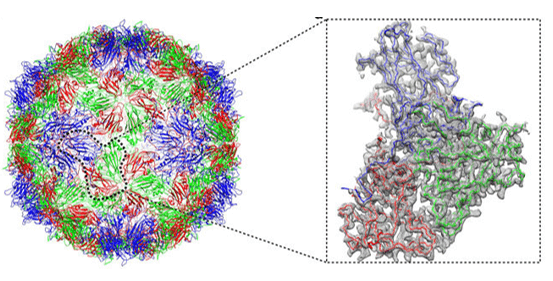
Case Study: Multi-Epitope Targeting TCRs and Structural Insights in Cancer Immunotherapy
This study identified that individual TCRs from successful melanoma TIL therapy can simultaneously recognize multiple tumor-associated antigens (TAAs) through shared structural motifs. Using combinatorial peptide libraries, proteomic searches, and the CANTiGEN platform, researchers revealed that single TCRs could bind epitopes from Melan A, BST2, and IMP2 presented by HLA-A*02:01.
High-resolution crystal structures of TCR-peptide-MHC complexes showed that cross-recognition was driven by conserved interaction hotspots, particularly at peptide positions 4 and 7, enabling a "molecular mimicry" mechanism across different TAAs. Multipronged TCRs demonstrated additive antigen recognition, enhanced tumor targeting, and resistance to antigen-loss escape mechanisms, outperforming monospecific TCRs.
Select Service
Related Reading
 Figure 5. Multi-Epitope Targeting by TCRs in Cancer Immunotherapy. (Dolton G, et al., 2023)
Combating Infectious Diseases
During the COVID-19 pandemic, rapid T cell epitope mapping SARS-CoV-2 played a crucial role:
- It revealed the breadth (covering Spike protein and other structural/non-structural proteins) and characteristics of T cell responses following natural infection and vaccination.
- It identified T cell epitopes cross-reactive between different coronaviruses.
- It pinpointed immunodominant epitopes in critical viral regions (especially conserved ones), providing vital information for addressing viral variants and designing next-generation broadly protective vaccines.
 Figure 6. Epitope Mapping Techniques for SARS-CoV-2 Research. Overview of in silico prediction and experimental mapping approaches to identify SARS-CoV-2 epitopes, supporting disease severity studies, diagnostics, vaccine, and therapeutic development. Methods include ELISA, ELISpot, Cryo-EM, and phage display analysis. (Hamed S M, et al., 2023)
Biotherapeutic Development and Safety
For protein-based drugs (e.g., monoclonal antibodies, therapeutic enzymes), predicting and experimentally mapping their potential T cell epitopes is a key step in assessing immunogenicity risk. If "hotspot" epitopes likely to trigger unwanted immune responses are identified, protein engineering techniques (e.g., site-directed mutagenesis, known as deimmunization) can be employed to reduce their binding to MHC or recognition by TCRs, thereby improving drug safety.
Immune Monitoring and Diagnostics
Tracking the dynamics of antigen-specific T cell responses during disease progression or following therapy (e.g., vaccination, immunotherapy). Developing T cell-based diagnostic tools for infectious diseases, autoimmune conditions, or cancer diagnosis and prognosis.
In summary, T cell epitope prediction and mapping are critical for advancing vaccine development, immunotherapy, infectious disease research, and biotherapeutic safety assessment. By integrating computational prediction with experimental validation, researchers can systematically identify immunodominant epitopes, optimize immune responses, and design more effective and safer immunological interventions.
To accelerate your research, Creative Biostructure offers comprehensive T cell epitope prediction and epitope mapping services, combining in silico screening with high-precision experimental validation. Contact us to learn how our expertise can support your project and drive your discoveries forward.
References
- Kurtz J R, Petersen H E, Frederick D R, et al. Vaccination with a single CD4 T cell peptide epitope from a Salmonella type III-secreted effector protein provides protection against lethal infection. Infection and Immunity. 2014, 82(6): 2424-2433. https://doi.org/10.1128/iai.00052-14
- Sanchez-Trincado J L, Gomez-Perosanz M, Reche P A. Fundamentals and methods for T‐and B‐cell epitope prediction. Journal of Immunology Research. 2017, 2017(1): 2680160. https://doi.org/10.1155/2017/2680160
- Hamed S M, Sakr M M, El-Housseiny G S, et al. State of the art in epitope mapping and opportunities in COVID-19. Future science OA. 2023, 9(1): FSO832. https://doi.org/10.2144/fsoa-2022-0048
- Peri A, Salomon N, Wolf Y, et al. The landscape of T cell antigens for cancer immunotherapy. Nature cancer. 2023, 4(7): 937-954. https://doi.org/10.1038/s43018-023-00588-x
- Dolton G, Rius C, Wall A, et al. Targeting of multiple tumor-associated antigens by individual T cell receptors during successful cancer immunotherapy. Cell. 2023, 186(16): 3333-3349. e27. https://doi.org/10.1016/j.cell.2023.06.020
- Tang X, Zhang W, Zhang Z. Developing T Cell Epitope-Based Vaccines Against Infection: Challenging but Worthwhile. Vaccines. 2025, 13(2): 135. https://doi.org/10.3390/vaccines13020135