Introduction: The Critical Role of Epitopes in Antibody Function
Antibodies are vital proteins of the immune system, responsible for recognizing and neutralizing pathogens, abnormal cells, or foreign substances with high specificity. This ability arises from their precise binding to specific regions on target molecules, known as antigens. The region on an antigen that an antibody binds to is called an antibody epitope, or antigenic determinant. Understanding antibody-epitope interactions is crucial for unraveling immune mechanisms, advancing diagnostics, and designing effective therapeutics.
Antibody epitope mapping identifies the specific binding site of an antibody on its target antigen. This complex yet valuable process reveals how antibodies recognize their targets and supports progress in biomedical research. This article explores the definition, significance, different types of epitopes, and the major mapping methods currently employed in antibody epitope mapping.
What Is an Antibody Epitope?
The epitope of an antibody is the specific region on an antigen that an antibody recognizes and binds to. It typically consists of a small group of amino acids, glycans, or lipids. This structure is recognized by the antibody's binding site, known as the paratope or Complementarity Determining Regions (CDRs). The interaction is highly specific and involves non-covalent forces such as hydrogen bonds, ionic bonds, van der Waals forces, and hydrophobic interactions.
Epitopes are classified into two main types based on their structure:
- Linear epitopes consist of consecutive amino acids in the antigen's primary sequence and remain recognizable even after protein denaturation.
- Conformational epitopes are formed by non-contiguous amino acids brought together through protein folding. Their recognition depends on the native 3D structure and is typically lost upon denaturation.
Distinguishing between these two types of epitopes is a prerequisite for selecting the appropriate antibody epitope mapping method. Most antibody epitopes on complex proteins are conformational, requiring techniques that can analyze the antigen's three-dimensional structure.
 Figure 1. Infographic illustrating the relationship between antibodies, epitopes, and paratopes, showing binding specificity in immune response. (Creative Biostructure)
Related Reading
Why is Antibody Epitope Mapping Crucial?
- Basic Research: By performing mapping of different epitopes on an antigen, we can gain a deeper understanding of protein structure-function relationships and reveal protein interaction interfaces.
- Drug Discovery and Development: Particularly the identification of therapeutic monoclonal antibody epitopes, which is vital for the development of antibody drugs. Understanding the precise binding site of an antibody helps predict its mechanism of action (e.g., whether it blocks ligand binding to a receptor or activates a signaling pathway), evaluate potential off-target effects, and guide antibody design and optimization to enhance affinity, specificity, and reduce immunogenicity. Monoclonal antibody epitope mapping is a key step in the therapeutic antibody development pipeline.
- Epitope-Based Vaccine Development: Identifying key epitopes (immunogenic epitopes) that can induce protective immune responses helps in designing more effective and safer subunit vaccines or peptide vaccines.
- Diagnostic Assay Development: Precise antibody epitope mapping aids in the development of high specificity and high sensitivity diagnostic antibodies for various detection methods such as ELISA, immunohistochemistry, and flow cytometry, avoiding false positives or negatives due to cross-reactivity.
- Intellectual Property Protection: For therapeutic antibodies, clearly defining the specific epitope they bind is a key aspect of patent applications and protection, clearly delineating the scope of the antibody's rights. Epitope information is frequently included in epitope claims to define the scope of protection and differentiate from prior art.
It can be said that precise antibody epitope mapping is a powerful engine driving antibody-related research and applications forward.
Key Methods and Technologies for Antibody Epitope Mapping
Antibody epitope mapping is a technology-intensive field, and various experimental and computational methods can be chosen depending on the required resolution, epitope type (linear or conformational), antigen characteristics, and available resources. Often, to obtain comprehensive and accurate information, researchers combine multiple techniques.
Experimental Antibody Epitope Mapping Techniques
A variety of experimental techniques are employed for antibody epitope mapping, each with its own principle, advantages, disadvantages, and suitability for different types of epitopes. Drawing from comprehensive comparisons, including studies on key therapeutic antibodies, these methods offer diverse approaches to understanding antibody-antigen interactions:
Peptide Scanning / Peptide Array (PepArr)
This is a common method for identifying linear epitopes. It involves synthesizing a series of short, overlapping peptides from the antigen sequence and testing their binding to the antibody, often using ELISA. While effective for some antigens and primarily targeting linear epitopes, studies show its success rate can be low, particularly for conformational epitopes. It generally provides peptide-level resolution and may reveal only partial epitopes.
Beyond peptide library ELISA, various ELISA formats can be employed for epitope mapping. This includes using truncated or mutated versions of the antigen, or competing different antibodies for binding to the antigen, to infer the location and relationships between epitopes. These methods are generally high-throughput and relatively low-cost, offering functional insights into binding.
 Figure 2. Linear Pepscan Mapping of SARS-CoV-2 RBD. Schematic of SARS-CoV-2 RBD Pepscan using overlapping 20-mer peptides to assess antibody reactivity. Hybridoma supernatants were first screened with peptide pools, followed by individual peptide analysis of immunodominant regions for hybridomas showing linear peptide binding. (Maghsood F, et al., 2022)
Site-Directed Mutagenesis (Alanine Scanning)
This protein engineering method involves substituting specific amino acid residues in the antigen (frequently to alanine) through genetic techniques and evaluating the impact on antibody binding. A significant reduction or loss of binding indicates that the mutated residue is crucial for the epitope. Alanine scanning (ALN) can provide residue-level resolution and helps identify critical amino acids within the epitope. However, caution is needed as these single-amino acid substitutions can sometimes inadvertently alter the overall structure of the antigen, potentially leading to the identification of false-positive epitope residues. It is relatively fast for cell surface antigens but more time-consuming for soluble proteins requiring purification. This method can be used to map both linear and conformational epitopes, though structural integrity is a key consideration.
 Figure 3. Epitope Mapping of prM Antibodies via Shotgun Mutagenesis. Fine epitope mapping of anti-prM monoclonal antibodies using DENV3 random mutagenesis and DENV4 alanine scanning libraries. Binding loss identified critical residues, shown as green spheres on the DENV2 prM/E protein structure. (A) MAbs not dependent on K26, (B) MAbs dependent on K26, (C) MAbs with key residues in both prM and E regions. (Smith S A, et al., 2016)
Phage Display
This technique involves creating libraries of bacteriophages that display random peptides or fragments of the antigen on their surface. These libraries are then screened with the antibody to identify phages displaying sequences that bind to the antibody. Sequencing the DNA of the binding phages reveals the peptide or protein fragment sequences that the antibody recognizes. This method is well-suited for identifying linear epitopes but can also be adapted to find conformational mimotopes.
 Figure 4. Schematic showing the phage display process for allergic peptide identification. Allergen genes are inserted into phages to express mimotope peptides. These phages are screened through biopanning using antigen-antibody binding. Non-binders are removed, while bound phages are amplified in E. coli. After multiple rounds of selection, specific clones are identified by serum assays, and peptide sequences are determined from the inserted DNA. (Zhou F, et al., 2021)
Domain Exchange (DomX)
Less granular than ALN, this method involves swapping structural domains between homologous proteins (e.g., between human and mouse versions of the same protein) and assessing changes in antibody binding. It is useful for identifying the general domain(s) involved in antibody binding, providing strand-level or domain-level resolution. While it can have a high success rate for identifying high-level binding regions, its application is limited to antigens with clearly defined, exchangeable domains, and results may require validation by other methods as it relies on natural sequence differences between the exchanged domains.
 Figure 5. Antibody Binding to Mouse/Human CTLA-4 Domain Exchange Mutants. Assessment of ipilimumab and tremelimumab binding to human CTLA-4, mouse CTLA-4, and domain-swapped CTLA-4 chimeras using cell-based ELISA. Ribbon diagrams show human domains in green and mouse domains in brown, illustrating species-specific binding patterns. (Dang X, et al., 2023)
Hydrogen-Deuterium Exchange Mass Spectrometry (HDX-MS)
This mass spectrometry-based technique measures the rate at which backbone amide hydrogens in the antigen exchange with deuterium from the solvent. When an antibody binds, it protects the epitope region from exchange, resulting in less deuterium uptake. HDX-MS is valuable for providing conformational information in a dynamic, soluble environment and is particularly suitable for studying conformational epitopes. It measures regions protected from solvent access ("protection") rather than direct binding points, meaning allosteric conformational changes induced by binding might also be identified as part of the "epitope." HDX-MS typically offers peptide-level resolution, which can sometimes lead to an overestimation of the actual epitope size.
 Figure 6. Epitope Mapping of ADAMTS13 by HDX-MS with scFv Binding. HDX-MS reveals antibody-binding sites in the MDTCS fragment of human ADAMTS13. (A) Peptides with reduced HDX upon scFv binding are shown in red, while unchanged peptides are black. (B) Protection factors and switchable residues are mapped at single-residue resolution. (C) Epitope visualized at both peptide and residue levels. (D) Spacer domain highlights slowed hydrogen exchange in the five-loop region, indicating the scFv binding epitope. (Casina V C, et al., 2015)
Hydroxyl Radical Footprinting (HRF)
Another mass spectrometry-based technique that utilizes hydroxyl radicals to chemically modify the side chains of accessible amino acid residues in the antigen. Antibody binding protects epitope residues from this modification. HRF offers residue-level resolution and, similar to HDX-MS, measures the protection of residues from solvent accessibility upon antibody binding rather than direct contact points. It can provide high-resolution insights but requires careful control of radical exposure time and often necessitates access to specialized equipment like synchrotron X-ray sources for optimal results.
 Figure 7. Schematic overview of the X-ray hydroxyl radical footprinting (HRF-MS) process. (A) Synchrotron radiation generates hydroxyl radicals that covalently label solvent-exposed residues on proteins. Antibody binding shields epitopes from oxidation. (B) Protease digestion followed by LC-MS/MS detects modified vs. unmodified peptides across exposure times. (C) Changes in solvent accessibility are quantified by plotting the fraction of unmodified peptide against exposure time to determine hydroxyl radical reactivity rate constants. (Kristensen L G, et al., 2024)
Chemical Cross-linking (XL)
This method involves using chemical linkers to covalently bridge amino acid residues that are in close spatial proximity within the antibody-antigen complex. Following digestion, mass spectrometry is used to identify the cross-linked peptides. XL can provide insights into the binding interface at approximately 3 Å resolution and has shown good alignment with X-ray crystallography data in comparative studies, often pinpointing residues within or close to the true epitope. It can also readily identify the corresponding residues on the antibody (paratope) that form the interaction surface. While it may sometimes "underestimate" the full epitope compared to crystallography due to linker specificity and size limitations, it provides clear evidence of spatial proximity and can reveal dynamic interactions not captured in static structures. It is relatively fast and requires less material than some other methods.
 Figure 8. Schematic of XL-MS for epitope mapping. Antibody and antigen are chemically cross-linked, followed by proteolytic digestion, then analyzed by liquid chromatography-tandem mass spectrometry (LC-MS/MS). The cross-linking data, combined with sequence information, helps identify interaction sites and epitope regions. (Ströbaek J, et al., 2025)
X-ray Crystallography and Cryo-Electron Microscopy (Cryo-EM)
These are powerful structural biology methods that provide atomic or near-atomic resolution views of the antibody-antigen complex. By determining the three-dimensional structure of the bound complex, the precise interaction interface and contributing residues can be directly visualized. These methods are considered the gold standard for identifying conformational epitopes at high resolution. However, they require successful crystallization of the complex (for X-ray) or preparation of suitable samples (for Cryo-EM), which can be challenging, time-consuming, and require significant expertise and specialized equipment.
 Figure 9. Polyclonal Epitope Mapping Using Electron Microscopy. Electron microscopy-based approach for mapping polyclonal antibody responses to an HIV-1 envelope vaccine. (Bianchi M, et al., 2018)
Select Service
Computational (In Silico) Epitope Prediction
Computational methods, often referred to as antibody epitope prediction, employ bioinformatic tools and algorithms to predict potential epitope regions in silico. Recent advancements, particularly in machine learning, have significantly enhanced these in silico prediction capabilities. These methods primarily fall into categories based on the input data used: those analyzing the antigen's primary amino acid sequence (sequence-based methods, often for linear epitopes) and those that require the antigen's three-dimensional structure (structure-based methods).
Sequence-Based vs Structure-Based Prediction Approaches
Sequence-based methods analyze patterns and properties within the amino acid sequence statistically correlated with known linear epitopes. Structure-based methods, leveraging the 3D conformation, evaluate surface characteristics like accessibility, shape, and electrostatic potential to predict both linear and, more importantly, conformational epitopes. Structure-based prediction methods are generally considered more powerful, especially for complex protein antigens where conformational epitopes are prevalent.
Benefits and Limitations of Computational Epitope Prediction
Computational prediction serves as a valuable preliminary step in antibody epitope mapping, providing researchers with hypotheses and guiding the design of more efficient experimental strategies by highlighting potential binding regions. However, in silico predictions are theoretical models and do not perfectly replicate the complex biological interactions. Therefore, they must always be validated through experimental mapping techniques to confirm the actual antibody binding sites.
Focused Insights: Monoclonal Antibody Epitope Mapping
Epitope mapping is critically important for therapeutic monoclonal antibodies due to the need for high specificity to ensure on-target binding and minimize side effects. Precisely mapping these epitopes is vital for understanding therapeutic mechanisms, optimizing structure, assessing cross-reactivity, and securing intellectual property.
Mapping mAb epitopes is challenging, especially for complex antigens like membrane proteins or those with multiple conformational states. While high-resolution structural methods like X-ray and Cryo-EM are often required, alternative methods (XL, HDX, HRF) provide valuable complementary data, revealing dynamic interactions that static structures might miss. For example, XL data for an antibody binding to CD27 suggested a potential dynamic interaction region not visible in crystallography.
The results from monoclonal antibody epitope mapping directly inform subsequent antibody design and engineering, serving as a key step in developing effective and safe antibody drugs.
Challenges and Considerations in Epitope Mapping
Despite continuous technological advancements, antibody epitope mapping still faces several challenges:
- Complexity of Conformational Epitopes: Conformational epitopes rely on the protein's overall three-dimensional structure, making their mapping more challenging than linear epitopes and requiring more complex experimental methods and data analysis.
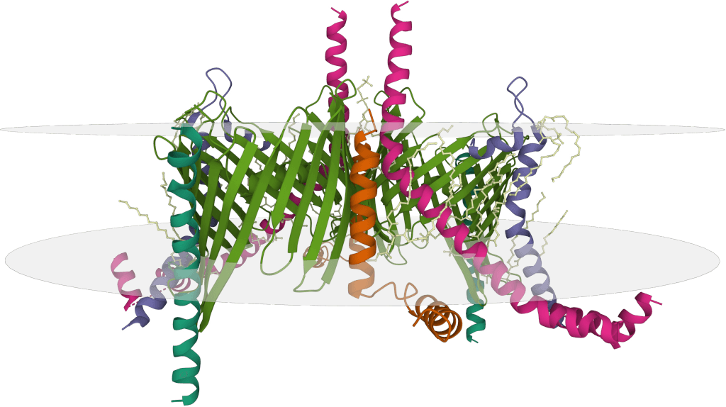
- Complex Antigen Challenges: For membrane proteins, multi-subunit complexes, or highly glycosylated antigens, their purification, folding, and formation of stable complexes with antibodies for structural analysis can be very difficult.
- Method Limitations: Each mapping method has its advantages and limitations and may only provide partial information. For example, peptide scanning is primarily suited for linear epitopes, while structural methods require successful sample preparation and resolution. HDX and HRF measure "protection" which might include allosteric effects.
- Data Analysis and Interpretation: Especially for large datasets from mass spectrometry or mutation scanning, their analysis and result interpretation require professional bioinformatics and structural biology expertise.
- Cost and Throughput: Some high-resolution methods (like structural determination) are costly and have low throughput, while others like peptide arrays also have limitations in success rate and resolution despite potentially higher throughput.
Choosing the Right Antibody Epitope Mapping Strategy
There is no single "one size fits all" antibody epitope mapping protocol. Selecting the most appropriate strategy requires comprehensive consideration of the following factors:
- Epitope Type: Does the target antibody recognize a linear epitope or a conformational epitope?
- Required Resolution: Do you need the location pinpointed to individual amino acid residues, or is knowing the general region sufficient?
- Antigen Characteristics: Is the antigen a protein? Is it a membrane protein or a soluble protein? What is its stability?
- Available Resources: Including sample quantity, purity, equipment conditions, and budget.
- Project Goals: What will the mapping results be used for (basic research, drug development, diagnostics, IP filing)? Different stages of drug development may benefit from different methods; crude mapping (DomX, Peptide Array, HDX) can be useful early for candidate prioritization and IP filing, while higher resolution methods (X-ray, HRF, XL, ALN) are important later for affinity maturation and understanding functional activity.
Often, researchers adopt a multi-technique integrated approach to obtain the most comprehensive epitope information.
Conclusion: Advancing R&D Through Epitope Insights
Antibody epitope mapping is a core process in immunology, structural biology, and drug development, revealing how antibodies interact with targets. This understanding is vital for designing new drugs, diagnostics, and studying immune responses. Technological advancements are making mapping more efficient for complex targets. While high-resolution methods like crystallography are key, complementary techniques provide valuable dynamic insights. Strategic use of these diverse methods is crucial for realizing antibodies' full potential.
Creative Biostructure specializes in antibody epitope mapping, offering comprehensive epitope mapping services integrating advanced techniques like HDX-MS, mutagenesis, peptide scanning, chemical cross-linking, and structural methods (X-ray, Cryo-EM). We tailor protocols for both linear and conformational epitopes, providing high-precision data and expert analysis. Contact us to gain detailed insights into antibody epitope binding, accelerating your research and drug development.
References
- Casina V C, Hu W, Mao J H, et al. High-resolution epitope mapping by HX MS reveals the pathogenic mechanism and a possible therapy for autoimmune TTP syndrome. Proceedings of the National Academy of Sciences. 2015, 112(31): 9620-9625. https://doi.org/10.1073/pnas.151256111
- Smith S A, Nivarthi U K, de Alwis R, et al. Dengue virus prM-specific human monoclonal antibodies with virus replication-enhancing properties recognize a single immunodominant antigenic site. Journal of virology. 2016, 90(2): 780-789. https://doi.org/10.1128/jvi.01805-15
- Bianchi M, Turner H L, Nogal B, et al. Electron-microscopy-based epitope mapping defines specificities of polyclonal antibodies elicited during HIV-1 BG505 envelope trimer immunization. Immunity. 2018, 49(2): 288-300. e8. https://doi.org/10.1016/j.immuni.2018.07.009
- Zhou F, He S, Sun H, et al. Advances in epitope mapping technologies for food protein allergens: A review. Trends in Food Science & Technology. 2021, 107: 226-239. https://doi.org/10.1016/j.tifs.2020.10.035
- Maghsood F, Amiri M M, Zarnani A H, et al. Epitope mapping of severe acute respiratory syndrome coronavirus 2 neutralizing receptor binding domain-specific monoclonal antibodies. Frontiers in Medicine. 2022, 9: 973036. https://doi.org/10.3389/fmed.2022.973036
- Dang X, Guelen L, Lutje Hulsik D, et al. Epitope mapping of monoclonal antibodies: a comprehensive comparison of different technologies. MAbs. Taylor & Francis, 2023, 15(1): 2285285. https://doi.org/10.1080/19420862.2023.2285285
- Jethva P N, Gross M L. Hydrogen deuterium exchange and other mass spectrometry-based approaches for epitope mapping. Frontiers in analytical science. 2023, 3: 1118749. https://doi.org/10.3389/frans.2023.1118749
- De Leon A J, Tjiam M C, Yu Y. B cell epitope mapping: The journey to better vaccines and therapeutic antibodies. Biochimica et Biophysica Acta (BBA)-General Subjects. 2024, 1868(10): 130674. https://doi.org/10.1016/j.bbagen.2024.130674
- Grewal S, Hegde N, Yanow S K. Integrating machine learning to advance epitope mapping. Frontiers in Immunology. 2024, 15: 1463931. https://doi.org/10.3389/fimmu.2024.1463931
- Kristensen L G, Gupta S, Chen Y, et al. Residue-Specific Epitope Mapping of the PD-1/Nivolumab Interaction Using X-ray Footprinting Mass Spectrometry. Antibodies. 2024, 13(3): 77. https://doi.org/10.3390/antib13030077
- Ströbaek J, Tang D, Gueto-Tettay C, et al. Epitope Mapping with Sidewinder: An XL-MS and Structural Modeling Approach. International Journal of Molecular Sciences. 2025, 26(4): 1488. https://doi.org/10.3390/ijms26041488