An epitope is a specific region on an antigen that is recognized and targeted by the immune system. Unlike the entire antigen, which may be a large and complex molecule, an epitope is a smaller segment that serves as the precise binding site for antibodies or immune cell receptors. This targeted recognition allows the immune system to accurately identify and respond to pathogens or abnormal cells, playing a critical role in immune defense.
A clear understanding of epitopes is essential not only for basic immunology but also for driving innovation in several critical areas. These include the development of effective vaccines, the creation of accurate diagnostic assays, and the design of targeted therapeutic antibodies. This foundational knowledge underpins techniques such as epitope mapping, which enables detailed exploration of these key molecular recognition sites.
What Exactly Constitutes an Epitope? Defining the Target Site
An epitope, frequently termed an antigenic determinant, represents the precise segment on an antigen molecule that an antibody or a T cell receptor recognizes and binds to. One can visualize an antigen as a complex structure; the epitope is akin to a specific keyhole within that structure, precisely designed to interact with a complementary key, the antibody or T cell receptor. This highly specific molecular interaction forms the fundamental basis of the adaptive immune response.
The intrinsic meaning of an epitope is deeply connected to its function as the molecular identifier that triggers an immune response. It is this distinct, interactive location on the antigen's surface that dictates which components of the immune system will engage with it.
Epitope vs Antigen: Distinguishing the Whole from the Part
It's important to clearly differentiate between an antigen and an epitope, as the terms are often used interchangeably but describe different entities. An antigen is typically a larger molecule capable of initiating an immune response. In contrast, an epitope is merely a specific, limited region or site on that larger antigen molecule.
A single antigen often carries multiple distinct epitopes. These different antigen epitopes can be recognized by various antibodies or immune cells, enabling a multi-pronged immune attack against a single pathogen or abnormal cell. This inherent multiplicity contributes significantly to the robustness and adaptability of the immune response. Grasping the difference between epitope and antigen is therefore key to appreciating the fine-tuned nature of immune recognition. The core difference between antigen and epitope lies in the scope of what is recognized: the antigen is the overall entity, while the epitope is the precise point of interaction for the immune system.
 Figure 1. Visual Guide to Antibody, Antigen, Epitope, Hapten, and Immunogen in Immunology. (Creative Biostructure)
Types of Epitopes: Structure and Immune Recognition
Epitopes exhibit considerable variation, primarily distinguished by their molecular architecture and the specific immune cell type that engages with them.
Characterization by Structural Arrangement
One primary means of classifying epitopes involves examining how the constituent residues or components are ordered within the antigen molecule's structure.
Linear (Continuous) Epitopes
These epitopes are defined by a direct sequence of connected building blocks, typically amino acids, forming an unbroken stretch along the antigen's chain-like structure. They are generally concise segments, often structured as helical formations roughly 9 to 12 amino acids in length. Linear epitopes are frequently encountered on molecules where their sequential composition is readily exposed, such as certain carbohydrates, fibrous proteins, and single-stranded nucleic acids. The binding interaction between an antibody and an antigen featuring a linear epitope is predominantly determined by the sequence of amino acids. For proteins, these epitopes may become accessible for antibody recognition only after the protein undergoes unfolding, a process like denaturation, which reveals internal linear sequences previously hidden within the folded structure.
Conformational (Discontinuous) Epitopes
In contrast, conformational epitopes possess a more intricate three-dimensional arrangement. They are formed by amino acid residues situated in different parts of the antigen's primary sequence that are brought into close spatial proximity through the molecule's folding into its specific native configuration. These epitopes are not linear; they are marked by breaks in the sequence but closeness in space. They are assembled from multiple disconnected segments that converge due to the antigen's natural folding. Such epitopes are typically larger than linear ones, often encompassing distinct groups of approximately 15 to 22 residues. Conformational epitopes are commonly located on the surface of most globular proteins and native nucleic acids, where the specific folded shape creates unique binding interfaces. Antibodies targeting a conformational epitope bind specifically to this precise three-dimensional structure. If the protein's folded structure is disrupted, for instance, by denaturation, this binding is usually lost because the essential spatial relationship between the residues forming the epitope is altered.
 Figure 2. Schematic of Linear and Conformational B-Cell Epitopes. (Ong Y C, et al., 2024)
Linear Epitope vs Conformational Epitope
| Feature | Linear Epitopes | Conformational Epitopes |
|---|---|---|
| Formation | Direct sequence of connected building blocks (e.g., amino acids). | Amino acid residues from different parts of the sequence brought together by 3D folding. |
| Residue Continuity | Continuous stretch of residues along the chain. | Discontinuous segments in the primary sequence, but spatially close. |
| Structural Complexity | Simpler, defined by sequence. | More intricate, defined by 3D arrangement. |
| Typical Size | Concise segments, roughly 9-12 amino acids. | Larger, often encompassing distinct groups of approximately 15-22 residues. |
| Location | Found on molecules with readily exposed sequential composition (e.g., certain carbohydrates, fibrous proteins, single-stranded nucleic acids). | Commonly located on the surface of most globular proteins and native nucleic acids. |
| Antibody Binding | Predominantly determined by amino acid sequence. May become accessible upon denaturation. | Determined by precise three-dimensional structure. Binding usually lost upon denaturation. |
Classification Based on Immune Cell Interaction
Another significant way to categorize epitopes is based on which type of immune cell primarily recognizes them: B lymphocytes or T lymphocytes.
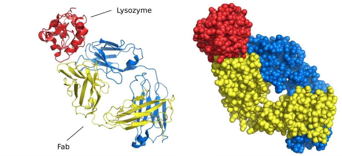
B-cell Epitopes
These are the regions on an antigen that are recognized by B lymphocytes through their surface-bound antibody molecules, known as B-cell receptors (BCRs). When an antibody engages with a protein, it targets a specific epitope, not the entire molecule. The site on the antibody or BCR that directly interacts with the epitope is termed the paratope, located at the tips of the antibody's antigen-binding regions (Fab). B-cell epitopes display considerable variability in their chemical nature and position on the antigen. Any solvent-accessible region on the antigen's surface can potentially serve as a B-cell epitope. These can range from small chemical substances to components of larger biological macromolecules like proteins, nucleic acids, lipids, and carbohydrates, although proteins are the most frequent category of antigen. Structurally, B-cell epitopes can be either linear (representing a smaller proportion, roughly 10%) or conformational (forming a large majority, around 90%). This prevalence of conformational B-cell epitopes highlights the crucial role of the antigen's native folded state for effective antibody recognition.
Epitope and Antibody/BCR Binding
The interaction between an epitope and its complementary antibody or BCR exemplifies molecular recognition driven by high specificity and strong binding. The epitope binding site on the antibody or BCR, known as the paratope, is structured with a shape and chemical properties that precisely complement those of the epitope. This epitope paratope interaction is often conceptualized as a lock and key mechanism, where the epitope serves as the lock and the paratope as its unique key. This precise fit allows the immune system to selectively target specific molecules on pathogens or altered cells, thereby minimizing unintended reactions against the body's own components. The strength of this binding, or affinity, is a critical factor in the efficacy of the immune response.
 Figure 3. Structure of B-Cell Receptor and Epitope Recognition. (Sanchez-Trincado J L, et al., 2017)
T-cell Epitopes
Unlike B cells, T lymphocytes do not interact directly with intact antigens. Instead, T cells recognize T-cell epitopes, which are short peptide fragments produced from the processing of protein antigens. These peptide fragments are displayed on the surface of specialized cells known as antigen-presenting cells (APCs), such as dendritic cells and macrophages, in conjunction with molecules from the major histocompatibility complex (MHC). The T cell receptor (TCR) on the T cell then specifically recognizes and binds to this particular peptide-MHC complex. T-cell epitopes are primarily derived from protein antigens. They are presented by two main types of MHC molecules: MHC Class I molecules, which present peptides to CD8+ cytotoxic T cells, and MHC Class II molecules, which present peptides to CD4+ helper T cells. This leads to the classification of CD8+ and CD4+ T-cell epitopes. While B-cell epitopes can be defined by both conformation and sequence, most T-cell epitopes are linear peptide sequences resulting from the intracellular breakdown of antigens.
 Figure 4. Recognition of T-Cell Epitopes by CD4⁺ and CD8⁺ T Cells. (Sanchez-Trincado J L, et al., 2017)
B Cell Epitope vs T Cell Epitope
| Feature | B-cell Epitopes | T-cell Epitopes |
|---|---|---|
| Recognizing Cell Type | B lymphocytes (B cells) | T lymphocytes (T cells) |
| Recognition Mechanism | Directly recognized by surface antibodies (BCRs). | Recognized as peptide fragments presented on MHC molecules by APCs. |
| Molecular Form | Intact antigen in native conformation. | Short peptide fragments. |
| MHC Requirement | No direct requirement for MHC presentation. | Requires presentation on MHC molecules (Class I or II). |
| Typical Source/Nature | Diverse: proteins, nucleic acids, lipids, carbohydrates; any exposed, solvent-accessible region. Proteins most frequent. | Primarily derived from protein antigens. |
| Structural Types | Can be linear (~10%) or conformational (~90%). | Mostly linear peptide sequences. |
| Binding Molecule | B-cell Receptor (BCR) / Antibody (via Paratope). | T-cell Receptor (TCR) binding to peptide-MHC complex. |
Significance of Epitopes in Biological Research and Beyond
The study and identification of epitopes are far from purely theoretical; they have profound implications for human health and the biotechnology sector. Pinpointing the specific epitopes that provoke an immune response is essential for several key applications:
- Vaccine Development: Identifying the crucial epitopes capable of eliciting protective immunity is a foundational step in creating effective vaccines. By focusing the immune response on these vital sites, vaccines can confer robust protection against infectious agents.
- Diagnostic Assay Design: Many diagnostic tests, such as Enzyme-Linked Immunosorbent Assays (ELISA), depend on the highly specific binding of antibodies to target epitopes present in patient samples, enabling the detection of diseases or infections.
- Therapeutic Antibody Engineering: Monoclonal antibodies used therapeutically, for example, in treating cancer or autoimmune disorders, are engineered to bind to specific epitopes on target cells or molecules, thereby modulating their function or marking them for elimination by the immune system.
- Epitope Tagging in Research: In molecular biology, short, well-characterized peptide sequences known as epitope tags are genetically fused to proteins of interest. Antibodies specific to these tags serve as versatile tools for detecting, purifying, or visualizing the tagged protein, even when antibodies against the untagged protein are unavailable.
Identifying Epitopes Using Epitope Mapping Technologies
Given the critical role epitopes play in immune recognition and their broad utility across research and development, the precise identification and characterization of these sites on an antigen is frequently essential. This is where the field of epitope mapping proves indispensable.
Epitope mapping encompasses a range of experimental techniques used to pinpoint the exact location and define the characteristics of epitopes on an antigen. This process is vital for:
- Determining if an epitope is linear or conformational.
- Identifying the specific amino acid or residue sequences involved in binding.
- Delineating the precise binding area of an antibody or T cell receptor on the antigen's three-dimensional structure.
Various techniques are employed depending on the type of epitope being studied:
- For mapping B-cell epitopes: Common methods include using overlapping synthetic peptides, alanine scanning mutagenesis, and structural techniques such as X-ray crystallography or cryo-electron microscopy.
- For mapping T-cell epitopes: Approaches often involve peptide binding assays, mass spectrometry-based MHC-peptide elution studies, and T-cell activation assays.
Precisely defining epitopes relevant to specific research or development goals is paramount. Epitope mapping provides the advanced tools necessary to achieve this detailed understanding, particularly given that a significant majority of B-cell epitopes are conformational and require sophisticated methods for their comprehensive characterization.
Select Service
Related Reading
- What Is Mass Spectrometry?
- Antigen-Antibody Complex Structure
- A Beginner's Guide to Antibody Epitope Mapping
- Overview of B Cell Epitope Prediction and Mapping
- Overview of T Cell Epitope Prediction and Mapping
- Difference Between Linear and Conformational Epitope Mapping
- Comparing Epitope Mapping Techniques
Conclusion
In summary, epitopes represent the vital, specific regions on antigens that serve as the targets for the adaptive immune system. Their highly specific interactions with antibodies and T cell receptors underpin immune specificity and the development of immunological memory. The diversity of epitopes, from their structural classification (linear versus conformational) to how different immune cells recognize them (B cell versus T cell), mirrors the inherent complexity of the immune response itself.
A deep understanding of epitopes is essential for advancing vaccines, diagnostics, and therapeutics that protect and improve human health. Precise identification and characterization of these sites through epitope mapping is a key step in immunology and biotechnology.
At Creative Biostructure, we offer professional epitope mapping services to accelerate your vaccine, diagnostic, and therapeutic development projects. Whether you're studying B-cell or T-cell epitopes, linear or conformational types, our advanced structural biology techniques ensure precise and actionable results. Contact us to discuss customized solutions tailored to your research needs.
References
- Sun J, Xu T, Wang S, et al. Does difference exist between epitope and non-epitope residues?. Immunome research, 2011, 7(3): 1.
- Luštrek M, Lorenz P, Kreutzer M, et al. Epitope predictions indicate the presence of two distinct types of epitope-antibody-reactivities determined by epitope profiling of intravenous immunoglobulins. PLoS One, 2013, 8(11): e78605. https://doi.org/10.1371/journal.pone.0078605
- Sanchez-Trincado J L, Gomez-Perosanz M, Reche P A. Fundamentals and methods for T‐and B‐cell epitope prediction. Journal of immunology research. 2017, 2017(1): 2680160. https://doi.org/10.1155/2017/2680160
- Hamed S M, Sakr M M, El-Housseiny G S, et al. State of the art in epitope mapping and opportunities in COVID-19. Future science OA. 2023, 9(1): FSO832. https://doi.org/10.2144/fsoa-2022-0048
- Ong Y C, Tejo B A, Yap W B. An Immunoinformatic Approach for Identifying and Designing Conserved Multi-Epitope Vaccines for Coronaviruses. Biomedicines. 2024, 12(11): 2530. https://doi.org/10.3390/biomedicines12112530