The rise of therapeutic antibodies has revolutionized medicine, enabling precise treatments for diseases such as cancer, autoimmune conditions, and infections. In addition to therapeutic use, antibodies are essential in diagnostics, research, and vaccine development. Their effectiveness depends on recognizing and binding to specific regions on antigens, known as epitopes, which is critical for guiding antibody design.
Analyzing how antibodies interact with these epitopes is a key step in development. Two commonly used and complementary approaches, epitope binning and epitope mapping, provide different insights. This article outlines their principles, applications, and differences to help researchers select the most suitable strategy for antibody characterization.
What Is an Epitope
Before delving into the characterization methods, it's crucial to reiterate the nature of the epitope. An epitope is the specific molecular structure on an antigen that an individual antibody recognizes and binds to. These recognition sites are diverse in nature and can be classified based on their composition and structural formation:
- Linear Epitopes: Composed of a continuous sequence of amino acids along the polypeptide chain of a protein antigen. Antibodies recognizing linear epitopes often bind even when the antigen is denatured, making them detectable in techniques like Western blotting.
- Conformational Epitopes: Formed by amino acid residues that are spatially close in the folded three-dimensional structure of the antigen but may be distant in the linear sequence. These epitopes rely on the correct folding of the protein, and antibodies binding conformational epitopes typically only recognize the native, biologically active form of the antigen.
Understanding whether an antibody binds to a linear or conformational epitope is a key piece of information, as it influences the choice of epitope mapping methods and provides insight into the potential function of the antibody, particularly if the native conformation is important for antigen activity.
Related Reading
Epitope Binning Classifying Antibodies by Binding Competition
High-throughput antibody discovery efforts commonly generate hundreds or even thousands of antibody candidates targeting the same antigen. Individually evaluating each of these candidates for functional activity or precisely mapping their binding location is often inefficient and time-consuming. Epitope binning provides a smart, high-throughput strategy to effectively group these antibodies based on their binding relationships, significantly streamlining the selection and characterization process.
What is Epitope Binning
Epitope binning is a technique used to categorize a panel of monoclonal antibodies (mAbs) based on whether they compete with one another for binding to the same or spatially overlapping regions (epitopes) on the target antigen. The core idea is that if two antibodies cannot simultaneously bind to the same antigen molecule, they likely recognize identical, overlapping, or very closely situated epitopes.
How Epitope Binning Works
The principle behind epitope binning is rooted in competitive binding assays. Typically, the target antigen is immobilized onto a solid support or sensor surface. Antibodies from the panel are then tested in pairwise combinations. A standard approach involves a "sandwich" or "pre-mix" assay format:
- Direct Binding: The antigen is captured on the surface.
- First Antibody Binding: A saturating concentration of a "first" antibody from the panel is flowed over the surface and allowed to bind to equilibrium.
- Second Antibody Binding: A "second" antibody (often labeled or detected via a secondary antibody) from the panel is then introduced.
If the second antibody can bind simultaneously with the first antibody, it indicates that the first antibody did not block its access to the antigen, suggesting they bind distinct epitopes. If the first antibody significantly reduces or completely prevents the binding of the second antibody, they are considered to compete for binding, implying recognition of the same or overlapping epitopes. This competitive behavior places them within the same "epitope bin."
This pairwise analysis is systematically performed for every possible combination of antibodies within the panel, creating a comprehensive matrix of binding relationships.
 Figure 1. Epitope Binning Workflow with Heatmap and Dendrogram Analysis. (Brooks B D, et al., 2020)
Technologies Enabling High-Throughput Epitope Binning
Efficiently performing the hundreds or thousands of pairwise competition experiments required for a typical antibody panel necessitates high-throughput label-free biosensing technologies. The most commonly used platforms utilize:
- Surface Plasmon Resonance (SPR): SPR instruments allow real-time monitoring of biomolecular interactions. By immobilizing the antigen on a sensor chip, the binding of antibodies and their competitive interactions can be measured directly by changes in the refractive index near the sensor surface. SPR platforms are designed for high throughput, enabling the analysis of large antibody panels against immobilized antigens.
- Biolayer Interferometry (BLI): BLI technology uses probes that dip into samples. Changes in the interference pattern of light reflected from layers on the probe tip indicate molecular binding. BLI systems are particularly well-suited for high-throughput screening and can analyze many interactions in parallel across multi-well plates.
Both SPR and BLI provide label-free, real-time data on antibody binding and competition kinetics, which is then analyzed computationally to determine blocking profiles and group antibodies into bins.
Select Service
Interpreting Epitope Binning Results
The output of a binning experiment is typically presented as a large matrix or "heat map," where rows represent the first antibodies and columns represent the second antibodies. The color or value in each cell indicates the extent of blocking observed between the corresponding antibody pair.
Sophisticated software analyzes this matrix to identify patterns of competition. Antibodies that exhibit similar blocking profiles against the entire panel are grouped together into "bins." Antibodies within the same bin are presumed to bind to the same or sterically overlapping epitopes. Antibodies in different bins are considered to bind to distinct epitopes. Network graphs are also commonly used to visualize the relationships between antibodies and the identified bins.
 Figure 2. Epitope binning of control anti-HEL antibodies. (A) Structural mapping shows five control antibodies bound across the HEL surface. (B) Sandwich assay using Octet for competitive binding analysis. (C) Heatmap summarizing blocking and sandwiching interactions among seven antibodies. (D) Network plot visualizing antibody blocking relationships and epitope bins. (Sivasubramanian A, et al., 2017)
Why Epitope Binning is Essential in Antibody Development
Epitope binning provides invaluable strategic information, particularly early in the antibody discovery pipeline:
- Efficient Lead Selection: It allows researchers to quickly understand the diversity of epitopes targeted by their antibody panel. Instead of pursuing every antibody clone, they can select one or a few representative antibodies from each distinct epitope bin for more in-depth functional characterization. This drastically reduces the number of candidates requiring further costly and time-consuming evaluation.
- Assessment of Epitope Diversity: Binning reveals how many distinct epitope regions on the antigen are recognized by the antibody library. High epitope diversity in a panel is often desirable, especially for therapeutic applications, as antibodies targeting different sites may have varied functional effects (e.g., blocking different protein-protein interactions).
- Identification of Functionally Relevant Epitopes: By correlating binning data with functional assay results (e.g., neutralization assays, cell-based signaling assays), researchers can identify which epitope bins are associated with desired biological activity. This helps prioritize antibodies targeting functionally relevant sites.
- Development of Complementary Antibody Pairs: Antibodies that fall into different bins, meaning they bind to distinct, non-overlapping epitopes, are ideal candidates for developing sensitive sandwich immunoassays (e.g., sandwich ELISA) used in diagnostics or pharmacokinetic studies.
- Strengthening Intellectual Property: Demonstrating that a proprietary panel of antibodies recognizes a diverse set of distinct or overlapping epitopes can provide stronger support for patent claims compared to simply claiming a large number of antibodies binding to the same antigen.
Limitations of Epitope Binning
While incredibly powerful for classifying antibody panels, it's important to note that epitope binning does not provide high-resolution structural details. It indicates if antibodies compete, suggesting spatial proximity or overlap, but it does not identify the exact amino acid residues or the precise structural features that constitute the epitope or the specific contact points between the antibody and the antigen at the molecular level.
Epitope Mapping Precisely Defining Antibody Binding Sites
Once a smaller, promising panel of antibody candidates has been identified and selected, often utilizing the insights gained from epitope binning regarding epitope diversity, the subsequent crucial step involves determining the exact location on the antigen where each antibody binds. This detailed identification process falls under the domain of epitope mapping.
What is Epitope Mapping
Epitope mapping is an experimental process aimed at pinpointing the specific amino acid residues, post-translational modifications, or distinct structural features on the surface of an antigen that constitute the binding site for a particular antibody. It moves beyond classifying antibodies by competition and focuses on identifying the precise molecular "footprint" of the antibody on its target.
The primary objective of epitope mapping is to gain detailed, often atomic-level, information about the antibody-antigen interface. This level of detail is essential for a deep understanding of the interaction and for downstream antibody engineering and development efforts.
Techniques for Epitope Mapping
Unlike the primarily competition-based methods of binning, epitope mapping employs a diverse range of biochemical, biophysical, and structural techniques, each providing different types of information about the epitope:
Mapping Linear Epitopes
- Peptide Scanning/Arrays: Overlapping synthetic peptides spanning the entire amino acid sequence of the antigen are synthesized and immobilized on a surface. The antibody is tested for binding to each peptide. Binding to a specific peptide or set of overlapping peptides identifies the linear epitope sequence.
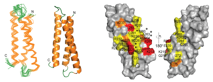
 Figure 3. Overview of multiplexed serological approaches for viral epitope mapping. Techniques include alanine scanning deep mutational scanning (top left), protein or peptide arrays analyzed via fluorescent antibody binding (top right), bacteriophage display libraries like Virscan with NGS readout (bottom right), and p-MHC multimers linked to DNA barcodes for epitope identification (bottom left). (Hu D, et al., 2023)
Mapping Conformational and Complex Epitopes (and sometimes linear)
- Site-Directed Mutagenesis: Key amino acid residues on the antigen (often guided by predictive algorithms or prior knowledge) are individually or combinatorially mutated. The effect of each mutation on antibody binding affinity is measured. Residues whose mutation significantly reduces or abolishes binding are identified as critical components of the epitope or essential for maintaining the epitope structure.
- Hydrogen-Deuterium Exchange Mass Spectrometry (HDX-MS): This powerful technique probes the solvent accessibility and dynamics of a protein. When an antibody binds its antigen, the region of the antigen contacted by the antibody becomes protected from exchange with deuterium in the solvent. By analyzing the rate of deuterium uptake in different peptide fragments of the antigen (via mass spectrometry) in the presence and absence of the antibody, the epitope region(s) can be localized, often providing information on conformational epitopes.
- Limited Proteolysis followed by Mass Spectrometry: The antibody-antigen complex is subjected to mild enzymatic digestion. The region of the antigen protected from cleavage by the bound antibody is then analyzed by mass spectrometry to identify the protected peptide fragments, which correspond to the epitope region.
- X-ray Crystallography: If the antibody-antigen complex can be crystallized, X-ray diffraction can provide a high-resolution 3D structure. This allows for the visualization of the precise amino acid residues on both the antibody and the antigen that make contact at the interaction interface, defining the epitope at the atomic level. This is the gold standard for mapping conformational epitopes.
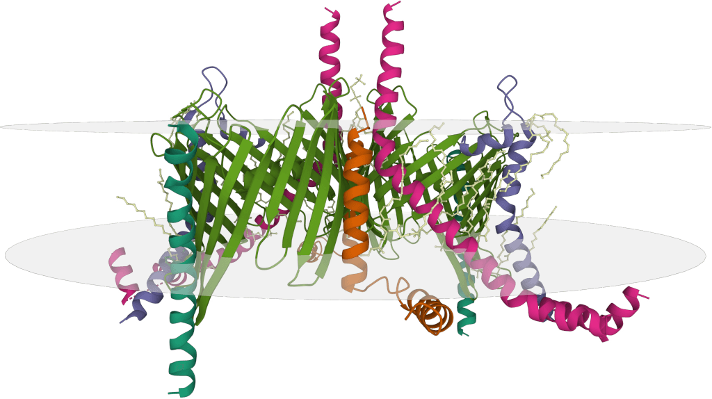
- Cryo-Electron Microscopy (Cryo-EM): For complexes that are difficult to crystallize, Cryo-EM can provide 3D structural information at various resolutions. While not always providing atomic resolution, it can often resolve the overall binding footprint and identify key regions involved in the interaction, particularly useful for larger antigens or membrane proteins.
Select Service
Computational and In silico Approaches
In addition to experimental methods, various computational tools and algorithms exist that can predict potential epitopes based on antigen sequence features (such as hydrophilicity or predicted surface accessibility) or existing structural data. While these in silico methods can be useful for generating hypotheses and guiding experimental design, they are predictive in nature and require rigorous experimental validation to confirm the actual epitope.
Mapping often involves applying a combination of these experimental techniques to build a comprehensive picture of the antibody's binding site.
Key Applications of Epitope Mapping
- Detailed Lead Candidate Characterization: It provides the in-depth molecular understanding required for regulatory submissions and patent filings. Knowing the exact epitope strengthens the description and claims associated with a therapeutic antibody.
- Mechanism of Action Elucidation: Mapping can definitively show if an antibody's epitope overlaps with a known functional site on the antigen, such as a receptor binding site, enzymatic active site, or protein-protein interaction interface. This provides direct evidence for the antibody's mechanism of action (e.g., whether it acts by blocking ligand binding or disrupting complex formation).
- Guiding Antibody Engineering: For efforts to improve antibody properties (e.g., affinity maturation, reduced immunogenicity, altered effector function), knowing the precise epitope ensures that modifications do not negatively impact critical binding interactions. It can also guide the design of antibody variants that target specific epitopes.
- Supporting Biosimilar Development: When developing a biosimilar antibody, demonstrating that it binds to the identical epitope as the reference product is a crucial part of establishing biosimilarity and requires rigorous mapping studies.
- Understanding Antibody Cross Reactivity: Precise epitope mapping can help explain why an antibody might cross-react with unintended targets by identifying shared structural features or sequences in the epitopes.
Challenges in Epitope Mapping
Despite its power, epitope mapping can present significant challenges. Mapping conformational epitopes, which depend on the antigen's three-dimensional structure, is generally more complex than mapping linear epitopes. Highly dynamic antigens or difficult-to-produce proteins, such as complex membrane proteins, can also pose hurdles. Structural methods like X-ray crystallography and Cryo-EM, while providing high resolution, can be technically demanding, time-consuming, and not always successful depending on the protein complex.
Epitope Binning vs Epitope Mapping
While both techniques are vital for antibody characterization, their fundamental approaches, the information they yield, and their typical applications differ significantly:
| Feature | Epitope Binning | Epitope Mapping |
|---|---|---|
| Core Question Asked | Which antibodies compete for binding? | Where on the antigen does the antibody bind? |
| Primary Information | Relative spatial relationship of epitopes | Precise molecular identity and location of the epitope |
| Resolution | Low to medium (groups of antibodies by region) | High (specific residues or structural elements) |
| Methodology | Competitive binding assays (typically label-free biosensors) | Diverse biochemical, biophysical, and structural methods |
| Application Focus | Antibody panel screening, diversity assessment | Lead candidate characterization, MOA, IP, engineering |
| Throughput Potential | High | Generally lower (varies by method) |
| Complexity | Relatively straightforward once platform is set up | Can be complex, requires specialized expertise & equipment |
| Output Example | Blocking heat map, antibody bin assignments | Peptide sequence, list of contact residues, 3D structure |
Epitope Binning and Epitope Mapping as Complementary Approaches
Instead of viewing them as competing approaches, epitope binning and epitope mapping are best seen as complementary techniques that provide different but equally valuable layers of information.
Epitope binning excels at efficiently sifting through a large pool of antibodies to understand the breadth of epitopes being targeted. By grouping antibodies that likely bind similar regions, binning drastically reduces the number of antibodies that need to undergo the more labor-intensive, high-resolution analysis of epitope mapping.
For example, if binning identifies five distinct epitope bins from a panel of 100 antibodies, researchers can select perhaps one or two representative antibodies from each bin. Mapping these 5-10 selected antibodies provides detailed information about five different epitope regions on the antigen targeted by the original panel. This strategic combination allows for both a broad overview of the antibody response (from binning) and detailed molecular insights into specific, functionally relevant interactions (from mapping).
Furthermore, binning data can sometimes provide initial clues that guide the design of mapping experiments. If antibodies in a certain bin exhibit a particular functional activity, mapping the epitopes for that bin becomes a high priority to understand the structural basis of that function.
Choosing the Right Epitope Characterization Strategy
Deciding whether to employ epitope binning, epitope mapping, or a combination depends heavily on the specific goals and stage of the antibody development project:
- Early Discovery with Large Panels: Epitope binning is usually the first step to classify antibodies, assess diversity, and select candidates for further analysis.
- Lead Candidate Selection and Optimization: Epitope mapping becomes essential to precisely define the binding site, understand the mechanism of action, and guide engineering efforts.
- Understanding Immunogenicity or Cross-reactivity: Mapping can help identify problematic epitopes.
- Biosimilar Development: Mapping the epitope of the reference product and the biosimilar is critical for comparability.
- Resource and Timeline Constraints: Binning offers a faster and often less expensive initial characterization compared to comprehensive mapping techniques like structural determination.
In a well-planned antibody development program, integrating both binning and mapping provides the most comprehensive understanding of antibody-antigen interactions, accelerating the selection of optimal candidates and supporting successful development.
At Creative Biostructure, we specialize in comprehensive antibody characterization solutions, including high-throughput epitope binning assay and high-resolution epitope mapping services. Whether you are screening large antibody libraries or validating lead candidates, our advanced platforms and structural biology expertise ensure accurate, reliable results tailored to your research needs. Contact us to discuss how we can support your therapeutic or diagnostic antibody development.
References
- Abdiche Y N, Miles A, Eckman J, et al. High-throughput epitope binning assays on label-free array-based biosensors can yield exquisite epitope discrimination that facilitates the selection of monoclonal antibodies with functional activity. PloS one. 2014, 9(3): e92451. https://doi.org/10.1371/journal.pone.0092451
- D Brooks B. The importance of epitope binning for biological drug discovery. Current drug discovery technologies. 2014, 11(2): 109-112.
- Yang G, Velgos S N, Boddapati S P, et al. Probing antibody‐antigen interactions. Antibodies for Infectious Diseases. 2015: 381-397. https://doi.org/10.1128/9781555817411.ch22
- Sivasubramanian A, Estep P, Lynaugh H, et al. Broad epitope coverage of a human in vitro antibody library. MAbs. Taylor & Francis, 2017, 9(1): 29-42. https://doi.org/10.1080/19420862.2016.1246096
- Chan B M, Badh A, Berry K A, et al. Flow cytometry-based epitope binning using competitive binding profiles for the characterization of monoclonal antibodies against cellular and soluble protein targets. SLAS DISCOVERY: Advancing Life Sciences R&D. 2018, 23(7): 613-623. https://doi.org/10.1177/2472555218774334
- Brooks B D, Closmore A, Yang J, et al. Characterizing epitope binding regions of entire antibody panels by combining experimental and computational analysis of antibody: antigen binding competition. Molecules. 2020, 25(16): 3659. https://doi.org/10.3390/molecules25163659
- Makowski E K, Wu L, Gupta P, et al. Discovery-stage identification of drug-like antibodies using emerging experimental and computational methods. MAbs. Taylor & Francis, 2021, 13(1): 1895540. https://doi.org/10.1080/19420862.2021.1895540
- Nagashima K, Mousa J J. Epitope binning of monoclonal and polyclonal antibodies by biolayer interferometry. Computational Vaccine Design. New York, NY: Springer US, 2023: 17-32.
- Hu D, Irving A T. Massively-multiplexed epitope mapping techniques for viral antigen discovery. Frontiers in Immunology. 2023, 14: 1192385. https://doi.org/10.3389/fimmu.2023.1192385