Cryo-EM for Ribosomes
The ribosome is a translational machinery that serves as the site of protein synthesis. It is made from complexes of ribosomal RNAs (rRNAs) and proteins which are arranged into two distinct components of different sizes. The ribosome is one of the most complex of protein/RNA, and obtaining the structure of the ribosome has remained a challenge due to the atom complexity of ribosomes and it is unstable because the aggregation and dissociation will happen when the environmental conditions and physiological state change.
Cryo electron microscopy (Cryo-EM) is an emerging and important methodology for structural biology. With the advance of microscope design, imaging hardware and image processing, etc., cryo-EM is increasingly becoming a mainstream technology for studying the structure of biological macromolecules at near-atomic resolution. It has been shown that Cryo-EM can solve many structures of the biological macromolecules while traditional crystallography cannot, especially for those that are difficult for crystallization, such as membrane protein, ribosome, and other large complexes.
At Creative Biostructure, we have the most advanced high-resolution electron microscopy equipment and a direct electron detector in combination with enhanced image processing, 3D reconstruction and atomic model building. Our platform has the ability to analyze ribosome structures from various species at the atomic level within their physiological environments.
The working process:
- Sample preparation: we have the advanced and mature technology of protein expression and purification to generate ribosomes (also can provide by customer).
- Vitrification of samples: the sample must be kept at cryogenic temperatures in order to minimize damage caused by the electron beam and result in high quality images.
- Image processing: Creative Biostructure utilizes the most advanced high-resolution electron microscopy and enhanced image processing to generate high resolution 2D images.
- 3D reconstruction and atomic model building: On the basis of high resolution 2D images to produce a 3D structural model by using computational procedures.
- Analysis of structure.
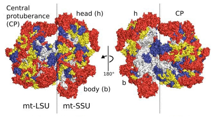 Figure 1. Overview of the human mitoribosome.
Creative Biostructure has a professional team for experimental design and Cryo-EM data analysis. We are dedicated to provide its customers with high quality, satisfying and excellent service.
Please feel free to contact us for a detailed quote.
Ordering Process

References
- Heena Khatter, Alexander G. Myasnikov, et al. Structure of the human 80S ribosome. Nature, 2015; 520:640-645.
- Alexey Amunts, Alan Brown, et al. The structure of the human mitochondrial ribosome. Sciences, 2015; 348:95-98.
- Eva Nogales. The development of cryo-EM into a mainstream structural biology technique. Nat Methods, 2016;13:24-27.