Liposome
Liposomes have been used as a good delivery carrier for many ion channel proteins and membrane transporters. Through a process of liposome separation, low-temperature sample preparation, and particle screening, three-dimensional (3D) structure reconstruction of the target protein can be obtained at ultra-high resolution by cryogenic electron microscopy (cryo-EM). Also, the size and shape of liposomes can be characterized by cryo-EM as it can directly visualize individual particles including their inner architecture.
The phospholipid bilayer structure of liposomes enables the target small molecules encapsulated in the bilayer. The intermediate region is the water phase, which can accommodate hydrophilic drugs. According to their different electric charges, common liposome materials can be divided into:
- Neutral phospholipid
- Phosphatidylcholine (PC)
- Phosphatidylethanolamine (PE)
- Sphingomyelin (SM)
- Negatively charged phospholipids (acidic phospholipids)
- Positively charged lipids
- Stearamide (SA)
- Cholesterol derivatives, such as CDBA and CTBBA
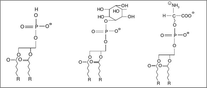 Figure 1. Structural formula of Phosphatidyl acid (PA), Phosphatidylinositol (PI) and Phosphatidylserine(PS)
Our team has rich experience in the processing and identification of liposome samples. We can perform cryo-EM structural analysis of membrane proteins embedded in liposomes and morphology analysis of liposomes.
If you are interested in our liposome particle morphology analysis service, please feel free to contact us. We are looking forward to cooperating with you.
Ordering Process


