Mapping Structure of Biological Drugs
Biological drugs are produced by living microorganisms, plants, or animal cells, and used in the prevention, diagnosis, or treatment of rheumatic arthritis, cancer and other diseases. Biological drugs are usually large biomolecules, such as monoclonal antibodies (mAbs) and interleukins. The replication of these protein molecules is a delicate task due to their structural complexity, and the potential safety risks. Several methods are available to characterize highly ordered structure of these drugs, i.e., NMR spectroscopy, X-ray crystallography, electron microscopy, mass spectrometry and functional assays. Creative Biostructure dedicated to aiding in biological drug design and discovery by its extraordinary biologists team specialized in protein structure. NMR is particularly useful in analysis of pharmaceuticals, screening weak-binding compounds for drug design.
Methyl group as NMR probe for mapping interaction between large biomolecules
The exquisite sensitivity of chemical shifts makes NMR spectroscopy an ideal method to study intermolecular interactions. However, this method was limited to small proteins. As recent approaches are developing, methyl group labeling and relaxation-optimized techniques (methyl TROSY) have overcome these restrictions and enabled studies of large molecules.
Methyl groups contain six amino acids, i.e. alanine, valine, leucine, isoleucine, methionine, and threonine. These methyl groups play as reporters of proper folding of a protein. Introduction of NMR-active methyl groups can be achieved with metabolic precursor. Methyl group labeling to specific residues could be obtained by chemical modification. By collecting methyl TROSY HMQC spectra, structural properties of binding surface of large molecular assemblies, such as antigen/antibody, pathogen molecule/receptors, kinase/inhibitor, can be studied.
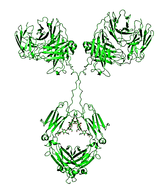
Investigating monoclonal antibody structure by 13C NMR at natural abundance
Generally, NMR is only applicable to biomolecules less than 30 KDa; for samples above 30 KDa, perdeuteration is required. If a high-resolution structure desired, stable isotope labeling is required (2H, 13C, 15N); the feasibility of applying NMR methods to stable isotope-labeled, protease-cleaved, mAb domains (Fab and Fc) has been demonstrated. However, isotopic labeling is not practical for analyzing drug products. Recent work reported application of 2D 13C methyl fingerprint method for structural mapping of an intact mAb at natural isotopic abundance (13C 1.1%). By combining of rapid acquisition and non-uniform sampling techniques, they were able to acquire a spectrum of 150 KDa mAb in a short time. Therefore, this method should serve as a precision tool for biopharmaceutical structural assessment.
Creative Biostructure is always enthusiastic about new techniques and willing to collaborate with research groups to develop methyl fingerprints NMR for biological drug discovery. Please contact us for more information!
Ordering Process

References
- Boštjan Japelj, Gregor Ilc, Jaka Marušič, Jure Senčar, Drago Kuzman & Janez Plavec (2016). Biosimilar structural comparability assessment by NMR: from small protein to monoclonal antibodies. Scientific reports. 6: 32201.
- Luke W. Arbogast, Robert G. Brinson, and John P. Marino, (2015). Mapping Monoclonal Antibody Structure by 2D 13C NMR at Natural Abundance. Analytical chemistry. 87(7): 3556-3561.
- Silke Wiesner and Remco Sprangers (2015). Methyl groups as NMR probes for biomolecular interactions. Current Opinion in Structural Biology.35: 60-67.