Inquiry Basket
| Your inquiry basket is currently empty. |
Aloe vera is rich in diverse bioactive compounds that contribute to its antioxidant, anti-inflammatory, and skin-supporting properties. Research highlights several key components and their mechanisms:
Aloe-derived exosomes are nanoscale extracellular vesicles (EVs) obtained from Aloe plant tissues. They act as natural carriers of Aloe-specific bioactive molecules and play roles in plant-based intercellular communication. Their main characteristics include:
These features make Aloe-derived exosomes a unique category of plant EVs with growing relevance in natural product and nanobiotechnology research.
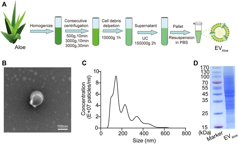 Isolation and Characterization of Aloe-Derived Extracellular Vesicle-Like Nanoparticles (EVAloe). (A) Schematic of EVAloe preparation involving differential centrifugation and sucrose gradient ultracentrifugation. (B) Transmission electron microscopy (TEM) image showing the morphology of EVAloe obtained from the 45% sucrose fraction. Scale bar: 100 nm. (C) Size distribution profile of EVAloe measured using nanoparticle tracking analysis (NTA). (D) SDS-PAGE analysis of EVAloe protein content using 10% polyacrylamide gel. (Zhou H, et al., 2023)
Aloe-derived exosomes offer several distinct advantages compared to synthetic or animal-derived extracellular vesicles, making them an attractive platform for advanced biomedical and biotechnology research:
In vitro studies have shown that Aloe exosomes do not induce cytotoxic effects in fibroblasts or endothelial cells, supporting their suitability for prolonged exposure. Being plant-derived, they carry no risk of animal pathogen transmission and do not require genetic modification.
Aloe exosomes have been shown to reprogram macrophages from pro-inflammatory (M1) to anti-inflammatory (M2) phenotypes, significantly reducing the expression of cytokines such as TNF-α and IL-6. In preclinical infection models, they alleviated tissue damage and cytokine storm responses. They have also demonstrated potential in promoting chronic wound repair by downregulating inflammatory genes (e.g., COX-2, iNOS) and enhancing fibroblast migration and proliferation.
Due to their nano-scale size (100-200 nm), Aloe exosomes can penetrate biological barriers such as the skin or intestinal mucosa. Their lipid bilayer structure protects encapsulated agents from enzymatic degradation, improving stability and circulation time. They have been used to deliver hydrophobic drugs, such as indocyanine green, for targeted photo-therapeutic studies, and inherently carry bioactive Aloe compounds like acemannan without chemical modification.
Aloe exosomes contain a variety of naturally functional molecules.
Cultivation of Aloe vera through optimized tissue culture protocols enables high propagation efficiency, with survival rates above 90%. Innovative extraction methods, including supercritical CO₂ and enzyme-assisted membrane filtration, enhance yield and preserve the integrity of heat-sensitive or bioactive components.
These combined advantages position Aloe-derived exosomes as a promising, plant-based extracellular vesicle platform for diverse research applications.
References
A study investigated aloe-derived nanovesicles (ADNVs) as a natural and stable nanocarrier system for drug delivery. Researchers successfully isolated ADNVs from aloe gel (gADNVs) and rind (rADNVs), with optimized conditions yielding monodispersed vesicles under 150 nm, suitable for systemic delivery. Compared to synthetic liposomes, gADNVs demonstrated superior structural stability, antioxidant capacity, and resistance to detergent-induced membrane disruption.
In drug-loading experiments, indocyanine green (ICG) was efficiently encapsulated in gADNVs, retaining over 90% of the active compound after 30 days of storage, compared to only 74% retention in ICG-loaded liposomes. Furthermore, ICG/gADNVs maintained phototherapeutic activity in vitro and inhibited melanoma tumor growth in vivo more effectively than control formulations. Fluorescence imaging revealed that gADNVs had enhanced transdermal penetration, reaching deeper skin layers than both free dye and liposome formulations.
The study highlights aloe-derived nanovesicles as promising plant-based carriers with high biocompatibility, long-term stability, and effective delivery performance, particularly suited for photosensitive or hydrophobic compounds.
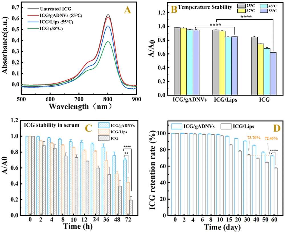 Figure 1. Evaluation of ICG Stability and Leakage in gADNVs Compared to Liposomes and Free ICG. (A) UV-vis spectra of ICG/gADNVs, ICG/Lips, and free ICG after 12 h at 55 °C. (B) Thermal stability comparison of ICG in different carriers. A/A₀ indicates the ratio of absorbance before and after heating. (C) Stability of ICG/gADNVs, ICG/Lips, and free ICG in serum. (D) Long-term retention rates of ICG in gADNVs and liposomes over 60 days.
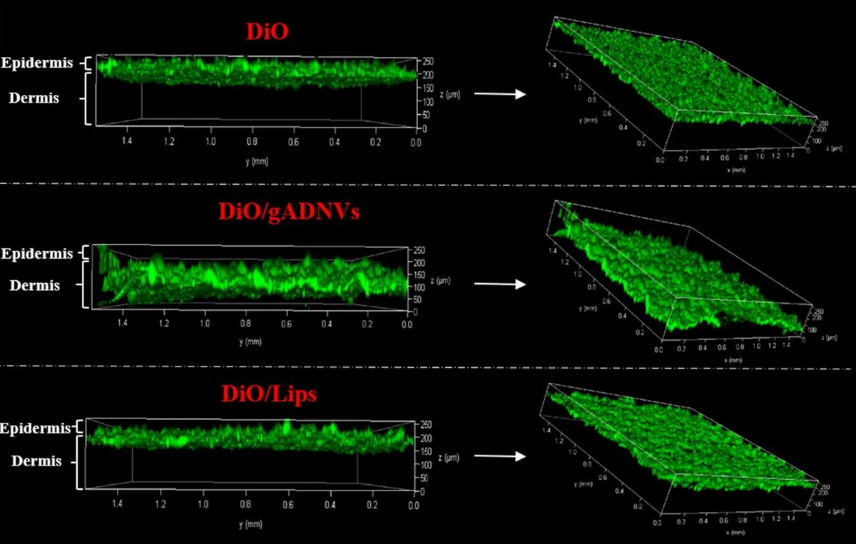 Figure 2. Enhanced Dermal Penetration of DiO via gADNVs Compared to Free DiO and Liposomes. Laser scanning confocal microscopy images showing the skin penetration of free DiO, DiO-loaded gADNVs, and DiO-loaded liposomes (DiO/Lips). DiO/gADNVs penetrated into the dermis with a broader distribution, while free DiO and DiO/Lips primarily remained in the epidermis, suggesting the potential of gADNVs as effective percutaneous drug carriers.
A study evaluated the potential of Aloe saponaria-derived extracellular vesicles (AS-EVs) for chronic wound healing. AS-EVs were isolated using a PEG-based precipitation method and confirmed to be spherical nanoparticles under 200 nm. Importantly, they showed no cytotoxicity in dermal fibroblasts, endothelial cells, or macrophages at concentrations up to 5 × 10⁹ particles/mL.
Functionally, AS-EVs reduced LPS-induced expression of inflammatory markers IL-6 and IL-1β by over 70%, indicating strong anti-inflammatory potential. They also promoted fibroblast proliferation (2.3-fold increase) and migration (over 3-fold), both essential for tissue repair. In tube formation assays, endothelial cells treated with AS-EVs formed longer capillary-like structures, up to 33% longer than controls, suggesting enhanced angiogenic activity.
These results support the role of Aloe-derived EVs as safe, plant-based vesicles with promising applications in inflammation modulation and tissue regeneration research.
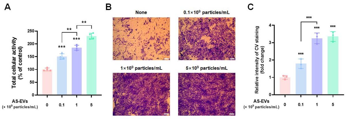 Figure 1. AS-EVs Promote Proliferation and Migration of Human Dermal Fibroblasts (HDFs). (A) WST-8 assay evaluating the viability of HDFs after 48 h incubation with AS-EVs in serum-free DMEM/F12. (B) Transwell migration assay showing enhanced migration of HDFs following 24 h AS-EVs treatment. Cells on the bottom side of the membrane were stained with crystal violet (CV) and fixed with 4% paraformaldehyde. Scale bar: 200 μm. (C) Quantification of migrated cells by measuring CV absorbance at 560 nm.
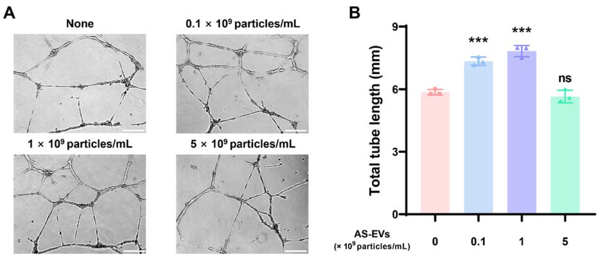 Figure 2. AS-EVs Enhance Capillary-Like Tube Formation in Human Umbilical Vein Endothelial Cells (HUVECs). (A) Representative images of capillary-like structures formed by HUVECs after 16 h of AS-EVs treatment on Matrigel in EGM-2 medium. Scale bar: 200 μm. (B) Quantification of total tube length using ImageJ.
References