Structured Illumination Microscopy Service
As an important tool in life science research, the fluorescence microscope has greatly promoted the development of life science. However, its resolution is limited, which affects the application in the field of subcellular life science. In recent years, with the development of science and technology, structured illumination microscopy (SIM) technology has greatly improved the resolution of the microscope system and effectively promoted the development of life science research.
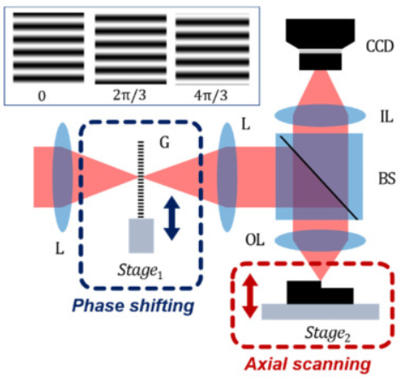 Figure 1. Typical structured illumination microscopy (SIM)
With a small light dose, low damage to the sample, and no restrictions on fluorescent dyes, SIM is suitable for the observation of dynamic processes in vivo.
SIM can get the structural information of specific parts of biological samples that cannot be obtained by conventional fluorescent microscopes. Three-dimensional SIM is used to obtain the super-resolution images of cytoplasmic filaments, mammalian nuclei, nuclear chromatin, nuclear fiber layer, nuclear pore complex, and other mammalian subcellular structure.
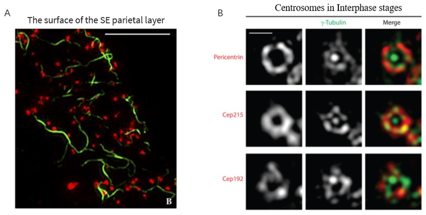 Figure 2. 3D-SIM image of subcellular structure
(A) The surface of the sieve elements parietal layer reveals strands of movement protein (MP)-decorated sieve element reticulum (green) associated with pore-plasmodesmata (red). (B) Centrosomes are shown for the interphase stage. γ-Tubulin (green) localization in interphase is shown relative to Pericentrin, Cep215 and Cep192 (red). Bar = 5 μm.
Creative Biostructure can provide subcellular structure analysis and microstructure analysis base on our electron microscope platform. Our services contain:
- Ultra-high resolution Fluorescence microscopy imaging service for tubulin in living cells (Green Fluorescence)
- Ultra-high resolution fluorescence microscopy imaging service for lysosomes in living cells (Deep Red Fluorescence)
- Ultra-high resolution fluorescence microscopy imaging service for lysosomes in living cells (Orange Fluorescence)
- Ultra-high resolution fluorescence microscopic imaging of mitochondria in living cells (Deep Red Fluorescence)
- Ultra-high resolution fluorescence microscopy imaging service for nuclei in living cells (Deep Red Fluorescence)
SIM is widely used in scientific research such as intracellular fine structure, accurate protein localization, dynamic observation of living cells, and so on. With our structured illumination microscopy platform, Creative Biostructure can provide sample preparation, data collection, and image analysis services to meet our customer’s requirements.
If you are interested in our structured illumination microscopy service, please feel free to contact us. We are looking forward to cooperating with you.
Ordering process

References
- Park H M, Joo K N. Motionless polarizing structured illumination microscopy. Sensors. 2021. 21(8): 2837.
- Fitzgibbon J, et al. Super-resolution imaging of plasmodesmata using three-dimensional structured illumination microscopy. Plant Physiology. 2010, 153(4): 1453-1463.
- Schermelleh L, et al. Subdiffraction multicolor imaging of the nuclear periphery with 3D structured illumination microscopy. Science. 2008. 320(5881): 1332-1336.
- Sonnen K F, et al. 3D-structured illumination microscopy provides novel insight into architecture of human centrosomes. Biology Open. 2012. 1(10): 965-976.
- York A G, et al. Resolution doubling in live, multicellular organisms via multifocal structured illumination microscopy. Nature Methods. 2012. 9(7): 749-754.


