Combinational Applications of Cryo-EM, X-ray Crystallography and NMR
Determination of the three-dimensional (3D) structures of biological molecules helps us better understand their biochemical features, and subsequently, their biological functions. Presently, X-ray crystallography, Nuclear Magnetic Resonance (NMR) spectroscopy, and Cryo-Electron Microscopy (Cryo-EM) are techniques that mainly used in the structural biology field, as they all can provide high-resolution structures of macromolecules. Following successful crystallization of samples, the X-ray crystallography technique allows identification of the 3D structures of both small molecules and large protein complexes. However, many proteins or protein complexes are difficult to crystallize due to solubility issues, the existence of flexible regions, and their high sensitivity to environmental changes. As a complementary approach, NMR can provide structural information about the disordered regions. Moreover, NMR also enables characterization of the molecule dynamics and their binding properties. However, NMR is typically used for the structural determination of proteins in the size range between 5 kDa to 25 kDa; and it becomes very difficult to apply for proteins larger than 50 kDa. In contrast to NMR, cryo-EM is not limited by molecule sizes – actually the bigger the size is, the better cry-EM will work. Moreover, unlike X-ray crystallography or NMR, the cry-EM technique does not require large amounts of sample to perform. Besides, cryo-EM allows determination of biomacromolecule structures in their native status as this method requires minimal sample preparation. Further, the sample structures can be directly visualized at a near-atomic resolution (better than 4 A˚) by an electronic detector.
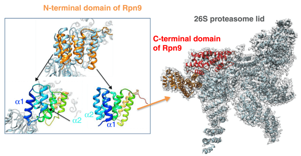 Fig. 1 Liquid-state NMR studies provide 3D structures for small components or binders of large complexes analyzed by Cryo-EM.
The combination of multiple methodologies such as Cryo-EM, X-ray crystallography and NMR allow researchers better investigate scientific questions by processing comprehensive analyses of the large, dynamic, macromolecular machineries in different organisms. At Creative Biostructure, we have our in-house X-ray Crystallography, EM and NMR platforms. Our excellent professional team will also provide intellectual and technical support for our customers, ensuring the quality and success of each project.
Creative Biostructure provides professional services in structural biology for the characterization of structure-based biological functions.
Please feel free to contact us for a detailed quote.
Ordering Process

References
- Philippe Cuniasse, Paulo Tavares, et al. Structures of biomolecular complexes by combination of NMR and cryoEM methods. Curr Opin Struct Biol. 2017; 43:104-113.
- Gabriel C Lander, Helen R Saibil, et al. Go hybrid: EM, crystallography, and beyond. Curr Opin Struct Biol. 2012; 22:627-635.
- Jeffrey Lengyel, Eric Hnath, et al. J Struct Funct Genomics. 2014; 15:117-124.


