Cryo-EM for Filaments
Cryogenic electron microscopy (cryo-EM) is a mainstream technology for studying the structure of biological specimens such as cells, viruses, and proteins. It can provide three-dimensional (3D) maps of biomolecules with near-atomic resolution. Cryo-EM overcomes the deficiency of transmission electron microscopy and can visually display the microstructures and sub-microstructures of cells and their components. It can be used to analyze the relationship between subcellar populations, or observe the ultrastructure of the surface or cross-section of filaments and ducts.
Filaments are long chains of proteins that are usually bound together to increase strength and hardness, such as microfilaments, microtubules, muscle filaments, etc. It is difficult to obtain the structure of the fiber by X-ray crystallography because of its large toughness and highly dynamic change. Our cryo-EM team provides high-resolution ultrastructural studies of filaments surfaces and cross-sections with rich sample information and accurate results.
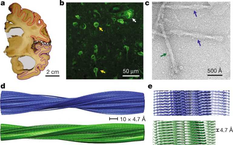 Figure 1. Structure of tau filaments from Alzheimer's brain (Fitzpatrick A W P, et al. 2017)
At present, with the continuous breakthrough in cryo-EM, pathology studies of amyloid fiber structure are also developing rapidly. More and more scientific research teams are now analyzing the fibrous or filament structures related to various diseases. With years of experience, we can also provide a high-resolution cryo-EM scanning service for your scientific research.
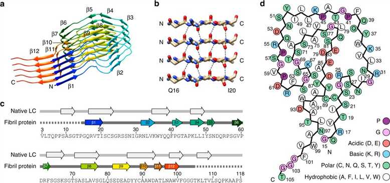 Figure 2. Cryo-EM structure of a light chain-derived amyloid fibril from a patient with systemic AL amyloidosis (Radamaker L, et al. 2019)
At Creative Biostructure, our service includes the preparation and purification of filaments, negative staining EM and image processing of Cryo-EM, provide the customer with the 3D reconstruction at a near atomic resolution, and finally provides a biologically insightful for understanding the biological progress.
Our cryo-EM team has rich experience in nanomaterial, subcellular organelles, filaments, and biological tissue or other EM samples. If you are interested in our filaments service, please feel free to contact us. We are looking forward to cooperating with you.
Ordering Process

References
- Fitzpatrick A W P, et al. Cryo-EM structures of tau filaments from Alzheimer’s disease. Nature. 2017, 547(7662): 185-190.
- Radamaker L, et al. Cryo-EM structure of a light chain-derived amyloid fibril from a patient with systemic AL amyloidosis. Nature Communications. 2019. 10(1): 1-8.