Electron Microscopy (EM) Platform
Electron microscopy (EM) is a very powerful technology for the ultrastructural study of macromolecules, cells and tissues. Experiencing major advances in recent years, EM is now able to achieve three-dimensional reconstruction of macromolecular assemblies at near-atomic resolution. The EM platform at Creative Biostructure is dedicated to provide service and scientific support to customers from both industry and academia for their research projects requiring high-resolution EM images of the biological samples of interest.
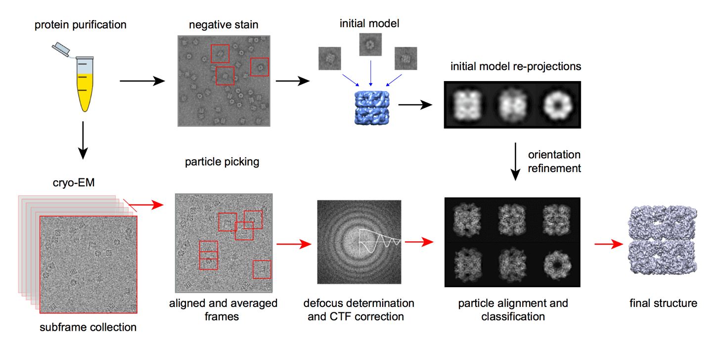 Figure 1. Workflow for single-particle reconstruction using EM.
Platform Equipment
Our platform is equipped with highly specialized instruments for scanning electron microscopy (SEM), transmission electron microscopy (TEM) and the auxiliary equipment required for cryo-EM.
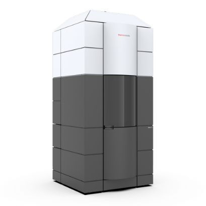
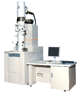
300 kV TEM (FEI)
This high-resolution electron microscope for 3D characterization of biological samples. Its cryo-based technology and stability allow a full range of semi-automated applications includes cryo-electron microscopy, single-particle analysis, dual-axis cellular tomography, and 2D electron crystallography of frozen-hydrated cell organelles and cells.
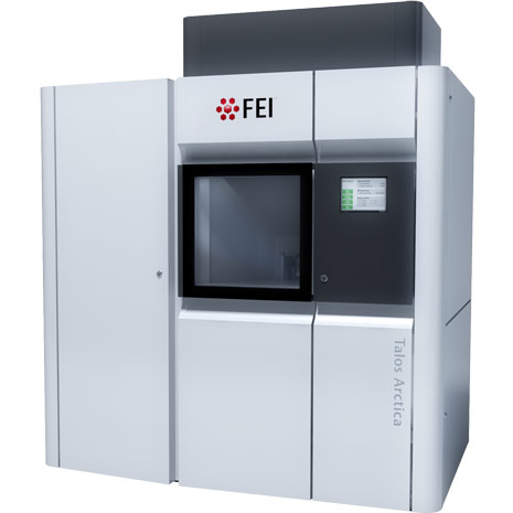
200 kV TEM (FEI)
This 200kV microscope combines high-resolution performance, high-tilt capability, and cryogenic autoloader capable of accommodating 12 samples at a time.
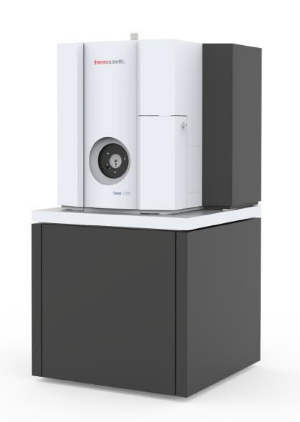
120 kV TEM (FEI)
This TEM is uniquely designed for performance across a wide range of applications, such as 2D and 3D imaging of cells, cell organelles, single-particle specimens, and soft materials, both at cryogenic (liquid nitrogen) and ambient temperatures.
Transmission Electron Microscope (JEOL JEM-2200FS)
This TEM combines a 200kV field emission gun (FEG) and in-column energy filter (Omega Filter), and uses a rotation-free image-forming optical system to produce a high-end, optimally configured TEM for energy filtered imagery and chemical analysis of specimens.
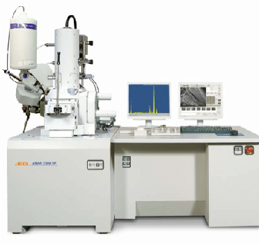
Scanning Electron Microscope (JEOL JSM-7001F)
This high-resolution SEM is equipped with a large specimen chamber that accommodates a wide variety of detectors simultaneously, including: multiple EDS, WDS, STEM, BSE, and CL. It is ideal for both imaging and analysis of nanostructures, and determining chemical composition of the sample through X-ray spectroscopy.
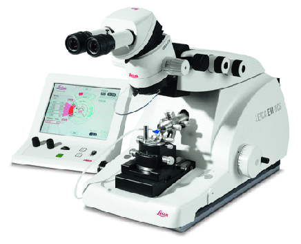
Ultramicrotome (Leica EM UC6)
This ultramicrotome, featuring an intuitive touchscreen control panel, allows fast and safer alignment of the programmable knife and specimen. With Leica’s AutoTrim, you can automatically trim a specimen to a predetermined level in the block face, ideal for high quality specimen preparation and morphometric studies.
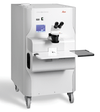
High Pressure Freezer (Leica EM HPM100)
This high pressure freezing system enables fast sample handling with excellent freezing results, ideal for vitrifying samples up to 200µm in thickness. Its unique 6 mm-diameter carrier system allows even more sample area to be frozen, compared with other high pressure freezing instrument.
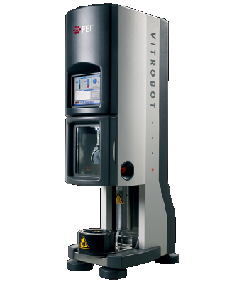
Vitrobot (FEI)
This system offers fully automated vitrification to provide fast, easy, reproducible sample preparation for high quality cryo-fixation at constant physical and mechanical conditions including temperature, relative humidity, blotting conditions and freezing velocity. The Vitrobot's controlled environmental technology prevents cooling and concentration of artifacts that are inevitable in other freezing protocols.
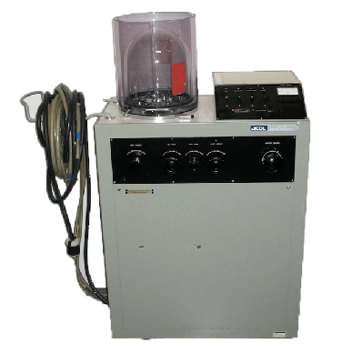
Vacuum Evaporator (JEOL JEE-4B)
This evaporator is a high-vacuum specimen preparation station for both SEM and TEM applications. It is particularly useful for the rapid deposition of carbon, gold and alloys on specimens prior to SEM or TEM imaging.
Sample Preparation
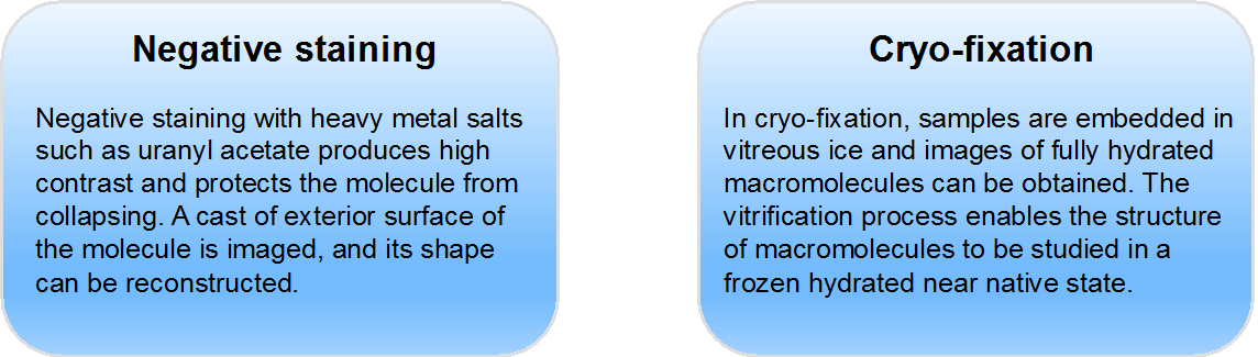
In our in-house EM platform, biological samples are typically prepared using negative staining or cryo-fixation. Negative staining usually has a lower signal-to-noise ratio, whereas cryo-fixation provides good preservation of native structural features of the sample.
Services
Our team at Creative Biostructure has expertise in preparing, imaging and interpreting a wide range of biological samples. We are proud to build this EM platform to support the following services that are customizable to meet the specific requirements of our customers:
- Scanning Electron Microscope Service
- Transmission Electron Microscope Service
- Cryo-electron microscopy (Cryo-EM)
Please feel free to contact us to discuss your project!
Ordering Process

References
- Cheng Y, Walz T (2009). “The advent of near-atomic resolution in single-particle electron microscopy”. Annu Rev Biochem 78:723-42.
- Thompson RF, et al (2016). “An introduction to sample preparation and imaging by cryo-electron microscopy for structural biology”. Methods 100:3-15.
- Carroni M, Saibil HR (2016). “Cryo electron microscopy to determine the structure of macromolecular complexes”. Methods 95:78-85.