Structural Research of Tumor Necrosis Factor (TNF) Receptor Superfamily
Members of the tumor necrosis factor receptor (TNFR) superfamily play critical roles in innate and adaptive immunity, influencing inflammation, lymphoid tissues, and T cell responses. Extensive evidence from human disease and experimental systems suggests that TNFR regulates the development of the immune system and the initiation of inflammatory responses for effective defense against viral and bacterial pathogens. Thus, the TNFR superfamily constitutes an essential molecular target for pharmacological intervention.
Advances in research on the TNFR superfamily
The TNFR superfamily consists of 29 transmembrane receptors, including extracellular domains responsible for ligand binding and intracellular domains that mediate the activation of signal transduction pathways. Researchers have classified TNFRs into two categories, activating receptors and death receptors (DRs). Most TNFRs are activating receptors, such as CD40 and TNFR2, which activate the nuclear factor κB (NF-κB) and mitogen-activated protein kinase (MAPK) pathways, while DRs consist of eight members, including TNFR1 and Fas, which have death domains (DDs) that mediate extrinsic signaling-induced cell death. Among them, TNFR1 is the most widely researched, which is capable of inducing both activation and death signaling pathways, thereby affecting cell metabolism, differentiation, and proliferation.
Structural insights into TNF-TNFR1 signaling
Researchers have demonstrated the biochemical and structural characteristics of the small, stable TNF-TNFR1 complex by X-ray crystallography, providing insights into the altered signaling upon binding of TNF to the receptor. The structure shows that TNF is a trimer, giving rise to three identical receptor-binding sites. TNFR1 and other TNFR superfamily members pre-aggregate before ligand binding and TNFR1 and TNFR2 signal after ligand binding. Both receptors have a similar structure to TNF's N-terminal extracellular domain (ECD), consisting of four cysteine-rich domains (CRDs), an α-helical transmembrane domain, and a cytoplasmic domain.
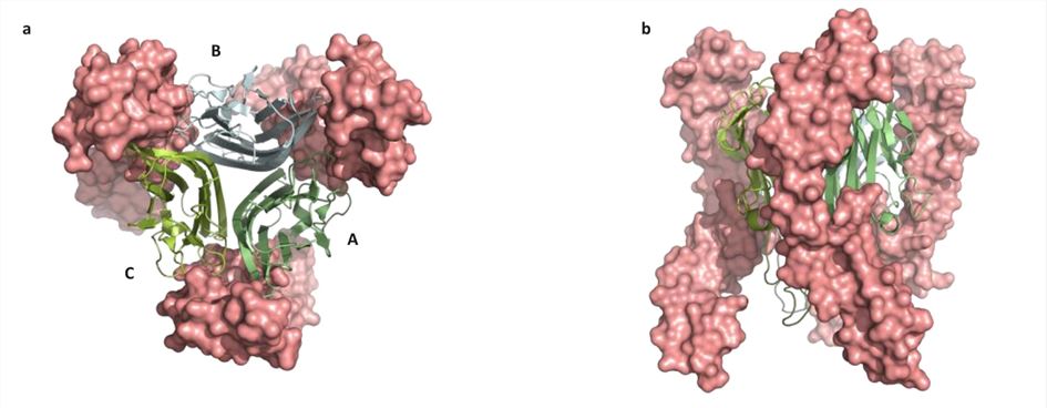 Figure 1. Top view (a) and side view (b) of TNF-TNFR1. (McMillan D, et al., 2021)
| Protein | Organism | Method | Resolution | PDB Entry ID |
| TNFR1 | Homo sapiens | SOLUTION NMR | / | 7K7A |
| DCR3 | Homo sapiens | X-ray diffraction | 2.901 Å | 3MHD |
| TL1A-DCR3 complex | Homo sapiens | X-ray diffraction | 2.951 Å | 3MI8 |
| DcR3-TL1A complex | Homo sapiens | X-ray diffraction | 2.45 Å | 3K51 |
| Carboxyl-terminal CARD-like domain TNFR-related death receptor-6 | Homo sapiens | SOLUTION NMR | / | 2DBH |
| 4-1BB and 4-1BBL | Homo sapiens | X-ray diffraction | 3.391 Å | 6A3V |
| 4-1BB/4-1BBL complex | Homo sapiens | X-ray diffraction | 2.13 Å | 6MGP |
| 4-1BB/4-1BBL complex | Homo sapiens | X-ray diffraction | 2.4 Å | 6BWV |
| 4-1BBL/4-1BB complex in C2 space group | Homo sapiens | X-ray diffraction | 2.7 Å | 6CPR |
| 4-1BB in P21212 space group | Mus musculus | X-ray diffraction | 2.6 Å | 5WJF |
| 4-1BB in P21 space group | Mus musculus | X-ray diffraction | 2.95 Å | 5WI8 |
| 4-1BB/4-1BBL complex | Mus musculus | X-ray diffraction | 2.65 Å | 6MKZ |
| Light mutant2 and dcr3 complex | Homo sapiens | X-ray diffraction | 2.78 Å | 4KGG |
| Light loop mutant in complex with dcr3 | Homo sapiens | X-ray diffraction | 2.27 Å | 4KGQ |
| Death receptor 6 | Homo sapiens | X-ray diffraction | 2.2 Å | 3QO4 |
| The S-SAD phased of the ectodomain of Death Receptor 6 (DR6) | Homo sapiens | X-ray diffraction | 2.96 Å | 3U3V |
| Preligand association of DR5 | Homo sapiens | SOLUTION NMR | / | 8DPX |
| GITR-GITRL complex | Mus musculus | X-ray diffraction | 3.302 Å | 7E57 |
| GITR (mGITR) with DTA-1 Fab fragment | Muromegalovirus G4 | Cryo-EM single particle analysis | 4.4 Å | 7RFP |
| Unliganded HVEM | Homo sapiens | X-ray diffraction | 1.9 Å | 5T2Q |
| RANK | Mus musculus | X-ray diffraction | 2.01 Å | 3ME4 |
| HVEM | Homo sapiens | X-ray diffraction | 2.251 Å | 4FHQ |
| Fab fragment of an anti-DR5 antibody bound to DR5 | Homo sapiens | X-ray diffraction | 3.2 Å | 4OD2 |
| OX40R (TNFRSF4) bound to Fab 3C8 | Homo sapiens | X-ray diffraction | 2.1 Å | 6OKM |
| OX40R (TNFRSF4) bound to Fab 1A7 | Homo sapiens | X-ray diffraction | 3.25 Å | 6OKN |
| Death Receptor 5 - Dimer of Trimer | Homo sapiens | SOLUTION NMR | / | 6NHW |
| YSd1 Fab bound to DR5 | Homo sapiens | X-ray diffraction | 3.35 Å | 1ZA3 |
| Death receptor 4 (DR4; TNFFRSF10A) bound to TRAIL (TNFSF10) | Homo sapiens | X-ray diffraction | 3 Å | 5CIR |
| FASL and DcR3 complex | Homo sapiens | X-ray diffraction | 2.5 Å | 4MSV |
| Fab BdF1 with Death Receptor 5 (DR5) | Homo sapiens | X-ray diffraction | 2.32 Å | 2H9G |
Table 1. Structural research of the tumor necrosis factor (TNF) receptor superfamily.
At Creative Biostructure, we offer various protein structure analysis services for the tumor necrosis factor receptor (TNFR). We have a team of highly skilled and experienced scientists with cutting-edge instruments and techniques such as X-ray crystallography, cryo-electron microscopy (cryo-EM), and NMR spectroscopy, who can provide customized solutions according to the client's specific requirements.
Through our services, clients can gain insight into the structural basis of TNFR and use this information to develop new therapies for autoimmune diseases. If you are interested in exploring the structure of the TNFR superfamily, please feel free to contact us to find out how we can help you in your scientific endeavors.
References
- McMillan D, et al. Structural insights into the disruption of TNF-TNFR1 signaling by small molecules stabilizing a distorted TNF. Nat Commun. 2021. 12(1): 582.
- Li J, et al. Structural basis of signal transduction in the TNF receptor superfamily. Adv Immunol. 2013. 119: 135-153.
- Puigserver, P. Signaling Transduction and Metabolomics. Hematology. 2018. 68-78.

