Types of Grids for Cryo-Electron Microscopy
Cryo-electron microscopy (cryo-EM) is one of the most used techniques in structural biology to analyze the three-dimensional structure of biological macromolecules. Although the sample preparation process for cryo-EM is simpler than that of another technique that is also very commonly used - X-ray crystallography. However, successfully preparing a sample suitable for high-resolution data collection still requires attention to many issues. Sample preparation is a key step in the whole technical route of cryo-EM. High-quality samples are the basis of everything; in order to carry the sample and make it possible to send it to the electron microscope for observation, the sample needs to be in contact with the carrier grid with a support film and fixed together by freezing. Therefore, choosing a suitable grid is the key to the success of cryo-EM.
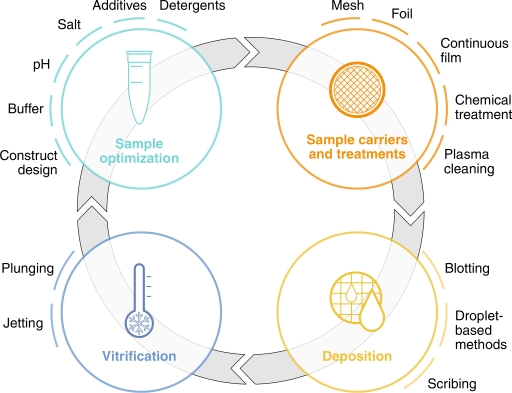 Figure 1. General overview of the stages involved in sample preparation for cryo-EM. (Weissenberger G, et al., 2021)
The carrier grid is a circular mesh with a diameter of 3 mm and consists of at least two parts: a mesh base and a support membrane. The mesh base is made of metal, usually copper, gold, or nickel, which plays the role of mechanical support, electron beam conduction, and heat dissipation. The support membrane is a perforated film with micron-sized holes placed on a mesh base.
With the development of cryo-EM technology, the importance of selecting the grid on the experimental results has been paid more and more attention. Different grids have different advantages and applications; the most used one is a copper grid with carbon film; the advanced choice is a gold grid with gold film; in addition, there are nickel-titanium alloy support film (ANTcryo) grids, etc.
- Copper Grid with Carbon Film
Copper grid with carbon film is the most used carrier mesh, which is a layer of 10-12nm thick carbon flat film attached to the surface of copper mesh; it has the most comprehensive application range, can provide a lower background signal, to obtain higher resolution cryo-EM data.
- Gold Grid with Gold Film
A gold mesh with a gold film is more conductive and simplifies the acquisition of cryo-EM data, even for challenging small molecules. During cryogenic imaging, traditional carbon films move, especially at the beginning of the scan. This movement blurs the image and reduces data quality. Furthermore, the porous support membrane made of metal will not shrink and deform before and after quick freezing, resulting in differences in results. It can improve the problem of adsorption of samples by the carbon support membrane, thereby improving the quality of the obtained data.
- Nickel-titanium Alloy Support Film (ANTcryo) Grid
The nickel-titanium alloy support film support grid can not only increase the conductivity, heat dissipation, and improve the particle penetration rate but also can be adapted to the routine alignment of the electron microscope and can also be directly applied to the current mature cryo-EM sample preparation and data collection. It has better conductivity and can effectively reduce sample drift caused by electron beam irradiation. Under the same conditions, a higher-resolution cryo-EM three-dimensional reconstruction result can be obtained. Therefore, the amorphous nickel-titanium microarray support film grid, as a new excellent performance grid for cryo-EM single-particle experiments, provides a new choice for most cryo-EM application researchers.
Leveraging our advanced cryo-EM technology, Creative Biostructure will choose the suitable carrier grid based on your research objectives and sample information, ensuring the acquisition of high-quality and repeatable data. Please don't hesitate to contact us for a detailed quote.
References
- Weissenberger G, et al. Understanding the invisible hands of sample preparation for cryo-EM. Nature Methods. 2021, 18(5): 463-471.
- Cheng H, et al. Dual-affinity graphene sheets for high-resolution cryo-electron microscopy. Journal of the American Chemical Society. 2023, 145(14): 8073-8081.

