Structural Research of Photosystem+Light-Harvesting Complex Supercomplex
During oxygen-producing photosynthesis, photosystems II (PSII) and I (PSI) operate in tandem for light-driven electron transport. Both photosystems contain a core and a peripheral antenna system. In plants, the peripheral antenna consists of the light-harvesting complex (LHC). LHCI and LHCII are connected to the PSI and PSII cores, respectively, to form the PS-LHC complex. Upon preferential PSII excitation, part of LHCII is phosphorylated and binds to PSI. The structural analysis of the PS-LHC complex is essential for revealing the energy transfer pathway between the antenna and the PSI core.
Structural analysis of PSI-LHCI supercomplexes
A total of three types of PSI-LHCI have been reported, two of which are supercomplexes with 10 Lhca proteins (PSI-10Lhca), while the other lacks the heterodimeric Lhca2/Lhca9 proteins (PSI-8Lhca). PSI-10Lhca and PSI-8Lhca structures are reconstructed by 3D cryo-electron microscopy (cryo-EM) at 2.9 Å and 3.1 Å resolution. The structures show that the four LHCI proteins (Lhca1-4) form two heterodimers, Lhca1/Lhca4 and Lhca2/Lhca3, which further form a heterotetramer that binds to the PSI core at the PsaF/PsaJ site, and the termini are stabilized by binding to PsaG and PsaK, respectively.
Progress in the structural research of PSI-LHCII supercomplexes
In plants, the transition from state 1 to state 2 is characterized by the formation of the characteristic PSI-LHCII supercomplex. The researchers analyzed the typical structure of PSI-LHCII by single-particle negative-staining electron microscopy (EM). where a single LHCII trimer binds to the PSI core on the PsaK side and attaches to the Lhca subunit. It was found that the transmembrane regions of PSI-LHCI and LHCII are not coplanar but form an angle of about 10°.
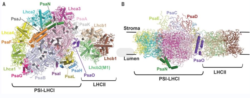 Figure 1. The maize PSI-LHCI-LHCII supercomplex. (Pan X, et al., 2018)
| Protein | Organism | Method | Resolution | PDB Entry ID |
| PSI-LHCI | Physcomitrium patens | Cryo-EM single particle analysis | 3.76 Å | 7KU5 |
| PSI-LHCR | Cyanidioschyzon merolae strain 10D | Cryo-EM single particle analysis | 3.63 Å | 5ZGB |
| M-LHCII and CP24 complexes in the stacked C2S2M2-type PSII-LHCII supercomplex | Pisum sativum | Cryo-EM single particle analysis | 3.6 Å | 5XNN |
| PSI-isiA supercomplex | Thermostichus vulcanus | Cryo-EM single particle analysis | 2.74 Å | 6K33 |
| PSII-FCP supercomplex | Chaetoceros gracilis | Cryo-EM single particle analysis | 3.02 Å | 6JLU |
| Photosystem I IsiA super-complex | Synechocystis sp. PCC 6803 | Cryo-EM single particle analysis | 3.48 Å | 6NWA |
| Photosystem I-IsiA supercomplex | Synechococcus elongatus PCC 7942 = FACHB-805 | Cryo-EM single particle analysis | 2.9 Å | 6KIG |
| Photosystem I-light harvesting complex I supercomplex | Pisum sativum | X-ray diffraction | 2.393 Å | 7DKZ |
| Photosystem I-LHCI super complex | Chlamydomonas reinhardtii | X-ray diffraction | 3.4 Å | 8H2U |
| Photosystem I-IsiA-flavodoxin supercomplex | Synechococcus elongatus PCC 7942 = FACHB-805 | Cryo-EM single particle analysis | 3.3 Å | 6KIF |
| Photosystem I supercomplex with light-harvesting complex I | Chlamydomonas reinhardtii | Cryo-EM single particle analysis | 2.9 Å | 6JO5 |
| C2S2M2N2-type PSII-LHCII | Chlamydomonas reinhardtii | Cryo-EM single particle analysis | 3.73 Å | 6KAF |
| Photosystem I supercomplex with light-harvesting complexes I and II | Zea mays | Cryo-EM single particle analysis | 3.3 Å | 5ZJI |
| State transition supercomplex PSI-LHCI-LHCII | Chlamydomonas reinhardtii | Cryo-EM single particle analysis | 2.84 Å | 7DZ7 |
| Photosystem I-LHCI-LHCII | Chlamydomonas reinhardtii | Cryo-EM single particle analysis | 3.42 Å | 7D0J |
| Photosystem I-LHCI-Lhca5 supercomplex | Hordeum vulgare subsp. spontaneum | Cryo-EM single particle analysis | 3.4 Å | 7EW6 |
| Photosystem I-LHCI-Lhca6 supercomplex | Hordeum vulgare | X-ray diffraction | 3.88 Å | 7EWK |
Table 1. Structural research of photosystem+light-harvesting complex supercomplex.
Creative Biostructure is dedicated to providing quality services for the research of membrane protein 3D structures. We use cryo-electron microscopy (cryo-EM) and X-ray crystallography to determine the structure of the photosystem+light-harvesting complex supercomplex, and provide clients with the best service and results through cutting-edge testing equipment and solutions. Our laboratory personnel have the expertise and proficiency to ensure accurate and reliable data. If you are interested in our services, please contact us for more details.
References
- Pan X, et al. Structure of the maize photosystem I supercomplex with light-harvesting complexes I and II. Science. 2018.360(6393):1109-1113.
- Crepin A, et al. Isolation and characterization of a large photosystem I-light-harvesting complex II supercomplex with an additional Lhca1-a4 dimer in Arabidopsis. Plant J. 2020.102(2):398-409.
- Yadav KN, et al. Supercomplexes of plant photosystem I with cytochrome b6f, light-harvesting complex II and NDH. Biochim Biophys Acta Bioenerg. 2017.1858(1):12-20.

