Structural Research of Bunyaviridae
The Bunyaviridae, now known as Bunyaviralesis, is a large family of viruses that is of great concern because many of its members cause serious diseases in humans and the outbreaks are increasing. Bunyaviridae consists of viruses with negative-sense RNA genomes that produce enveloped spherical virions 80-120 nm in diameter. In recent years, researchers have found that all Bunyavirus genomes consist of three segments (S, M, and L) encapsidated in a single ribonucleoprotein complex encoding glycoproteins Gc and Gn, nucleoprotein N, and viral polymerase L.
Classification of Bunyaviridae

- Arenaviridae
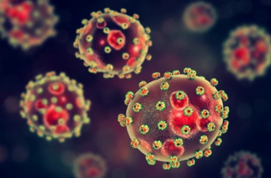
It presents as a spherical shape with an envelope. Diameter from 60 to 300 nm.
Arenaviruses are usually transmitted by rodents, and these rodent species are natural reservoirs of the virus. Arenaviruses can cause various viral hemorrhagic fevers and lymphocytic choriomeningitis (LCM).

- Hantaviridae
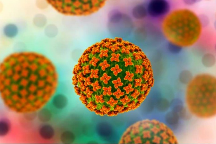
It presents as a spherical shape with an envelope. Diameter from 80 to 120nm.
Transmitted primarily through mice and rats, it causes viral hemorrhagic fever with renal syndrome (HFRS) and hantavirus pulmonary syndrome (HPS).

- Nairoviridae
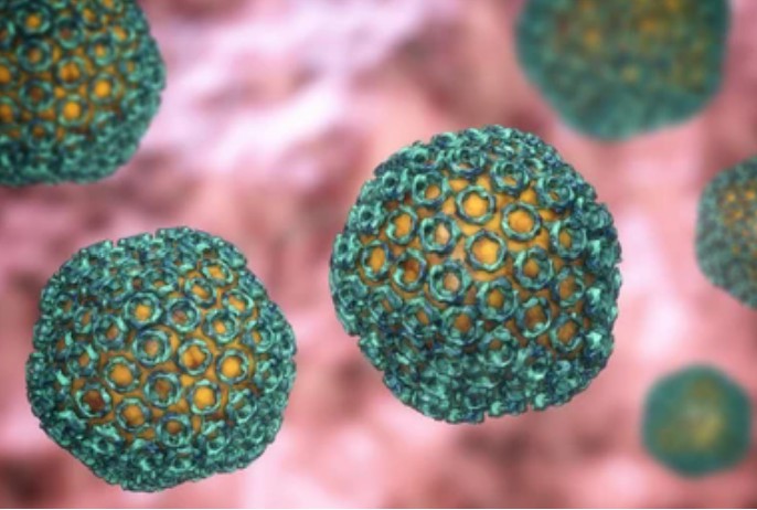
It presents as a spherical shape with an envelope. Diameter from 80 to 120nm.
Most nairoviruses are transmitted by ticks. The most essential nairoviruses are the Crimean-Congo hemorrhagic fever viruses, which cause Crimean-Congo hemorrhagic fever (CCHF).

- Phenuiviridae
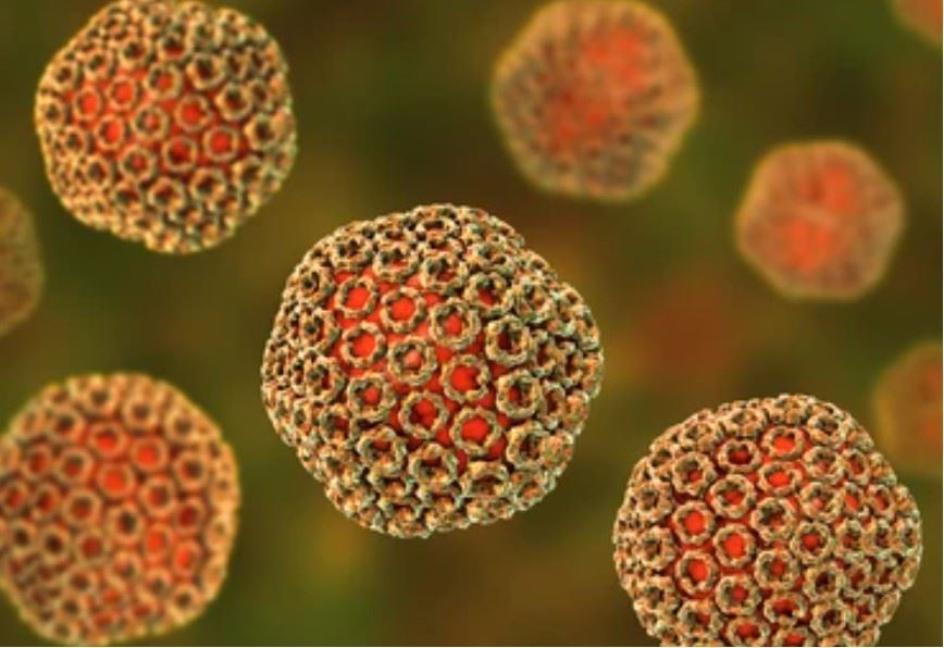
Enveloped, spherical. Diameter from 80 to 120 nm. Glycoproteins on the envelope surface are arranged on an icosahedral lattice with a symmetry of T=12.
Ruminants, camels, humans, and mosquitoes are natural hosts for Phleboviruses, which cause diseases (e.g., Rift Valley fever).
Structural characterization of Bunyaviruses is difficult using electron microscopy due to their glycoprotein-covered envelope and polymorphic structure. Therefore, Creative Biostructure offers advanced cryo-electron tomography (cryo-ET) technology, a powerful tool for analyzing complex viral structures. We can provide molecular resolution 3D images of undisturbed viral particles, allowing in situ visualization of structures.
In addition, we offer a comprehensive range of virus-like particles (VLPs) products. Our cutting-edge products are designed to satisfy the requirements of researchers, vaccine developers, and diagnostic professionals. Our virus-like particles are self-assembling structures that mimic the morphology and surface characteristics of viruses, making them ideal tools for research on virus entry, immunology, and vaccine development.
| Cat No. | Product Name | Virus Name | Source | Composition |
| CBS-V557 | Bunyamwera virus VLP (NSm Proteins) | Bunyamwera virus | Mammalian cell recombinant | NSm |
| CBS-V656 | Hantaan virus VLP (GP; NP; CD40L Proteins) | Hantaan virus | Mammalian cell recombinant | GP; NP; CD40L |
| CBS-V764 | Chimeric SBV & OROV VLP (N(SBV); L(SBV); M(OROV) Proteins) | Schmallenberg virus; Oropouche virus | Mammalian cell recombinant | N(SBV); L(SBV); M(OROV) |
| Explore All Bunyaviridae Virus-like Particle Products | ||||
Choose Creative Biostructure as a trusted partner to advance virology research. We specialize in the field of structural biology, and our scientists have expertise in the structural analysis of biomolecules and are proficient in X-ray crystallography and cryo-electron microscopy (cryo-EM). We offer contract services for the identification and characterization of viral particles according to clients' requirements. If you are interested in our services and products, please contact us for more information.
References
- Overby AK, et al. Insights into bunyavirus architecture from electron cryotomography of Uukuniemi virus. Proc Natl Acad Sci U S A. 2008. 105(7): 2375-2379.
- Ter Horst S, et al. Structural and functional similarities in bunyaviruses: Perspectives for pan-bunya antivirals. Rev Med Virol. 2019. 29(3): e2039.