Structural Research of Filoviridae
The Filoviridae are a family of single-stranded retroviruses, the members of which typically produce filamentous, enveloped viral particles that cause some of the most serious and fatal viral diseases in humans and non-human primates. The best known of these members are the Ebola virus (EBOV) and Marburg virus (MARV), which can cause systemic infections associated with severe hemorrhagic fever and high mortality. Research has found that the filovirus ribonucleoprotein complex, called nucleocapsid, plays a critical role in viral invasion. In recent years, researchers have used cryo-electron microscopy (cryo-EM) to resolve critical features of EBOV and MARV nucleocapsid structures. The structural analyses have revealed the possible mechanisms of nucleocapsid assembly, providing potential targets for antifiloviral drug design.
Morphological Features of Filovirus Particles
Research has shown that filoviruses have a diameter of about 80nm, and purified virions may be as long as 790-970nm, with a complex structure that includes the nucleocapsid, envelope, polymerase complex, and matrix. The shape of the virus is filamentous, U-shaped, 6-shaped, or round, and the surface has tumor-like projections scattered throughout the lipid bilayer. The helical capsid is located at the central axis of the filamentous particle and houses the host-derived lipid bilayer envelope and the VP40 inner layer lining the underside of the envelope. The VP40 forms a regular layer beneath the viral envelope. Viral particles on the surface of the viral particle are covered by GPs, forming surface spikes approximately 7nm in diameter.
Structure of the EBOV and MARV Nucleocapsids
The helical core coat of filoviruses was found to be similar to that of other non-segmented negative-sense RNA viruses, acting as a template for vRNA synthesis. The filovirus capsid consists of five VPs, NP, VP35, VP24, VP30, and L. At the initial stage of capsid formation, the NP encapsidates vRNA to form a helical NP-RNA complex, which interacts with the VP35, VP30, and L to form a functional capsid capable of vRNA synthesis. Subsequently, VP24 binds to the functional nucleocapsid in preparation for viral particle incorporation. Using cryo-electron tomography (cryo-ET), the researchers determined the overall structure of the mature EBOV and MARV nucleocapsid at a resolution of approximately 10 Å. Both the EBOV and MARV capsids show the left-handed helical core of the NP-RNA complex with a prominent portion consisting of VP35 and VP24.
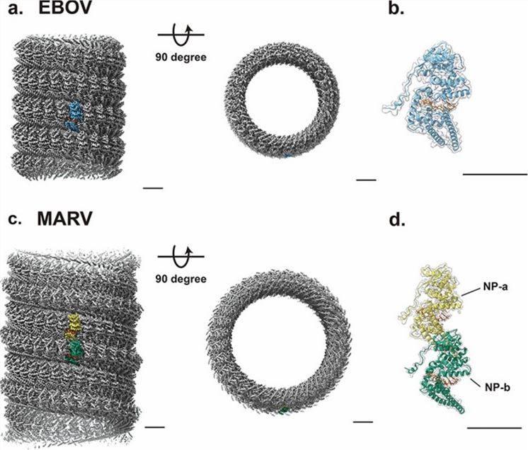 Figure 1. Cryo-electron microscopy of EBOV and MARV NP-RNA complexes. (Hu S, Noda T., 2023)
Creative Biostructure is a leading supplier of virus-like particles (VLPs) products for immediate global delivery. The VLPs are multi-protein structures that mimic the organization and conformation of real, natural viruses, and are non-infectious and safe to handle. Our VLP products help clients understand the three-dimensional structure of viruses are widely used in vaccine and antiviral drug development, and also have essential applications in other biotechnologies, including viral-like vectors for gene therapy, antibody screening, immunogen production, and ligand binding assays.
| Cat No. | Product Name | Virus Name | Source | Composition |
| CBS-V061 | Ebola virus VLP (GP, NP, VP 40 Proteins) | Ebola virus | Insect cell recombinant | GP, NP, and VP40 |
| CBS-V064 | Sudan virus VLP (GP, NP, VP40 Proteins) | Sudan virus | Insect cell recombinant | GP, NP, and VP40 |
| CBS-V102 | Ebola VLP (Z glycoprotein) | Ebola virus | Mammalian cell recombinant | Z glycoprotein |
| CBS-V670 | Marburg virus VLP (VP40; GP; NP Proteins) | Marburg virus | Mammalian cell recombinant | VP40; GP; NP |
| Explore All Filoviridae Virus-like Particle Products | ||||
As a cutting-edge biological company providing viral structure analysis services and virus-like particles (VLPs) products, Creative Biostructure is the best choice for every researcher in viral structural biology. We demonstrate professional services and experienced operations in data collection, image selection, and classification throughout the entire process of cryo-electron microscopy (cryo-EM) characterization.
We are committed to understanding the unique requirements and goals of each client and providing tailored solutions. If you have any questions, please feel free to contact us. We will provide you with the most accurate and detailed insights into viral architecture and assembly for the design and development of vaccines and antiviral drugs.
References
- Hu S, Noda T. Filovirus helical nucleocapsid structures. Microscopy (Oxf). 2023. 72(3): 178-190.
- Hu S, et al. Cryoelectron microscopic structure of the nucleoprotein-RNA complex of the European filovirus, Lloviu virus. PNAS Nexus. 2023. 2(4): pgad120.
- Kuhn JH, et al. ICTV Virus Taxonomy Profile: Filoviridae. J Gen Virol. 2019. 100(6): 911-912.

