Structural Research of Dihydroorotate Dehydrogenases (DHODH)
Dihydroorotate dehydrogenases (DHODH) proteins from different organisms have been divided into two classes. The first class includes the cytoplasmic family, while the second class includes membrane associated DHODH class 2, to which human DHODH belongs, allows for the reoxidation of FMN using ubiquinone and the presence of a highly variable N-terminal extension responsible for enzyme binding to the inner mitochondrial membrane. the N-terminal extension folds into a separate structural domain with two α-helices. It is located at the top of the catalytic C-terminal structural domain near the FMN binding site and contributes to the formation of a tunnel-like pocket. Some class 2 DHODH inhibitors were found to bind within this tunnel, which led to the suggestion that ubiquinone might also bind there. Human DHODH has two structural domains, the α/β-barrel structural domain containing the active site and the α-helical structural domain that forms the tunnel opening to the active site. Both inhibitors share a common binding site in this channel, and differences in the binding region determine drug sensitivity or resistance.
DHODH inhibitors have been recommended for the treatment of rheumatoid arthritis, psoriasis, autoimmune diseases, malaria parasites, and bacterial and fungal infections. The characterization of the structure of the complex between DHODH and inhibitors can provide new insights for the development of inhibitors.
The flavinase dihydroorotic acid dehydrogenase (DHOD, EC 1.3.99.11) catalyzes the oxidation of dihydrowhey ester to whey acid, which is the fourth step in the biosynthesis of UMP pyrimidine from scratch. The first crystal structure of rat type 2 dihydroDHOD has been determined in the complex of its two inhibitors, brequina and atorvaquinone. A unique feature of type 2 DHODs is their N-terminal extension, which folds into a separate domain containing two α helices. This domain serves as the binding site for two inhibitors, while respiratory quinones serve as the second substrate for two types of DHOD.
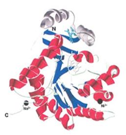 Figure 1. DHODR-breq, DHODR in complex with brequinar (light blue). (HANSEN M, et al., 2004)
| Protein | Organism | Method | Resolution | PDB Entry ID |
| Dihydroorotate Dehydrogenase | Escherichia coli | X-ray diffraction | 1.70 Å | 1J79 |
| Dihydroorotate Dehydrogenase | Escherichia coli | X-ray diffraction | 1.90 Å | 1XGE |
| Dihydroorotate Dehydrogenase in complex with atovaquone (expressed in E. coli) | Rattus rattus | X-ray diffraction | 2.30 Å | 1UUM |
| DHO in complex with brequinar | Rattus rattus | X-ray diffraction | 2.40 Å | 1UUO |
| Dihydroorotate Dehydrogenase, apo form | Homo sapiens | X-ray diffraction | 3.00 Å | 2PRM |
| DHODH in complex with brequinar analogue | Homo sapiens | X-ray diffraction | 2.40 Å | 2PRH |
| DHODH in complex with "a novel inhibitor" | Homo sapiens | X-ray diffraction | 3.00 Å | 2PRL |
| Dihydroorotate Dehydrogenase with triazolopyrimidine-based inhibitor DSM1 (expressed in E. coli) | Plasmodium falciparum 3D7 | X-ray diffraction | 2.00 Å | 3I65 |
| Dihydroorotate Dehydrogenase with bound triazolopyrimidine-based inhibitor DSM2 | Plasmodium falciparum 3D7 | X-ray diffraction | 2.40 Å | 3I68 |
| Dihydroorotate Dehydrogenase with bound triazolopyrimidine-based inhibitor DSM74 | Plasmodium falciparum 3D7 | X-ray diffraction | 2.50 Å | 3I6R |
Table 1. Structural Research of Dihydroorotate Dehydrogenases (DHODH).
Creative Biostructure employs leading-edge X-ray crystallography technology, and we use state-of-the-art instrumentation and have extensive laboratory experience to provide our clients with high-quality 3D structural analysis of membrane proteins. We analyze and process samples to obtain high-resolution crystallographic images of membrane proteins that reveal the internal structure and localization of functional regions. Our goal is to provide our clients with accurate, reliable and high-quality structural analysis services to enhance their research progress in related fields. If you are interested in learning more about our protein structural analysis services, please contact us for more information.
References
- THODEN J B, et al. Molecular structure of dihydroorotase: A paradigm for catalysis through the use of a binuclear metal center. Biochemistry, 2001, 40(24): 6989–6997.
- LEE M, et al. Dihydroorotase from escherichia coli: Loop movement and cooperativity between subunits. Journal of Molecular Biology, 2005, 348(3): 523–533.
- HANSEN M, et al. Inhibitor binding in a class 2 dihydroorotate dehydrogenase causes variations in the membrane-associated N-terminal domain. Protein Science, 2004, 13(4): 1031–1042.
- WALSE B, et al. The structures of human dihydroorotate dehydrogenase with and without inhibitor reveal conformational flexibility in the inhibitor and substrate binding sites. Biochemistry, 2008, 47(34): 8929–8936.
- DENG X, et al. Structural plasticity of malaria dihydroorotate dehydrogenase allows selective binding of diverse chemical scaffolds. Journal of Biological Chemistry, 2009, 284(39): 26999–27009.

