Super-Resolution Microscopy Service
Creative Biostructure has access to advanced super-resolution light microscopy facility allowing images to be taken with a higher resolution than the diffraction limit. Super-resolution microscopy is a form of light microscopy, which can be applied to observe many biological structures not resolvable in conventional fluorescence microscopy. For example, new advances in this technique now give them the ability to image three-dimensional (3D) structures, measure interactions by multicolor colocalization, and record dynamic processes in living cells at the nanometer scale.
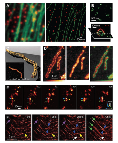 Figure 1. Examples of super-resolution images of biological samples.
Several far-field imaging methods are known to extend the diffraction-limited resolution to smaller values:
- Deterministic super-resolution: The most commonly used emitters in biological microscopy, fluorophores, show a nonlinear response to excitation, and this nonlinear response can be exploited to enhance resolution, such as STED, GSD, RESOLFT and SSIM.
- Stochastic super-resolution: The chemical complexity of many molecular light sources gives them a complex temporal behavior, which can be used to make several close-by fluorophores emit light at separate times and thereby become resolvable in time, such as Super-resolution optical fluctuation imaging (SOFI) and all single-molecule localization methods (SMLM) such as SPDM, SPDMphymod, PALM, FPALM, STORM and dSTORM.
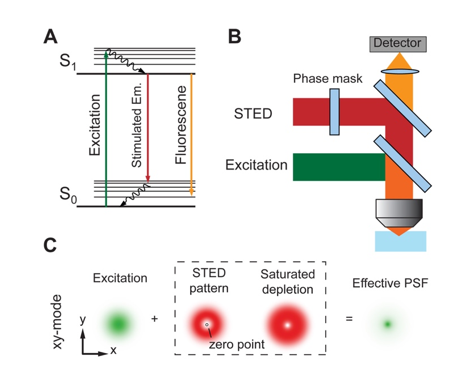 Figure 2. The principle of STED microscopy.
Applications in Super-Resolution Microscopy technology:
- Three-dimensional (3D) imaging
- Multicolor imaging
- Live cell imaging
Super-resolution fluorescence microscopy has shown great promise and ability in studying biological structures and processes at the cellular to macromolecular scale. Images obtained from these new super-resolution approaches will enable scientists to directly visualize biological samples at the nanometer scale and will complement the insights obtained through traditional molecular and cell biology approaches, significantly expanding our understanding of molecular interactions and dynamic processes in living systems. Please feel free to contact us for a detailed quote.
Ordering Process

References
- Neice, A. Methods and Limitations of Subwavelength Imaging. Advances in Imaging and Electron Physics. 2010. 163: 117-140.
- W.E. Moerner plenary presentation: Single-molecule spectroscopy, imaging, and photocontrol -- foundations for super-resolution microscopy. SPIE Newsroom. March 2015.
- Bo Huang, Mark Bate, et al. Super resolution fluorescence microscopy. Annu Rev Biochem. 2009; 78: 993–1016.


