Diagnostic Electron Microscopy Service
The electron microscope (EM) is used as an important analytical method in the physical, chemical and biological sciences. Compared with light microscope, scanning electron microscopy (SEM) and transmission electron microscope (TEM) use electrons to image tissues at much greater magnification, so EM becomes an essential diagnostic tool to screen human tissues at high magnification and allows for visualization of details within cells. As a powerful tool for visualizing the tissues, cells, virus, nanomaterials as well as other biopsy, TEM is particularly important in tumor pathology, neuropathology, kidney disease, and so on.
When combined with other methods, such as immunohistochemical and molecular genetic techniques, EM exhibits even greater impact on sample analysis and visualization, especially for understanding the relationships of the structure and function at the subcellular level. For example, light microscopy and immunofluorescence techniques are not able to provide confident result in the diagnosis of ependymoma due to that not all ependymomas are immunoreactive to epithelial membrane antigen (Figure 1A). EM, on the other hand, delivers ultrastructure of tissues and shows a well-developed cellular junctions, such as desmosomes, microvilli, and cilia, thereby allowing for direct diagnosis of ependymoma (Figure 1B).
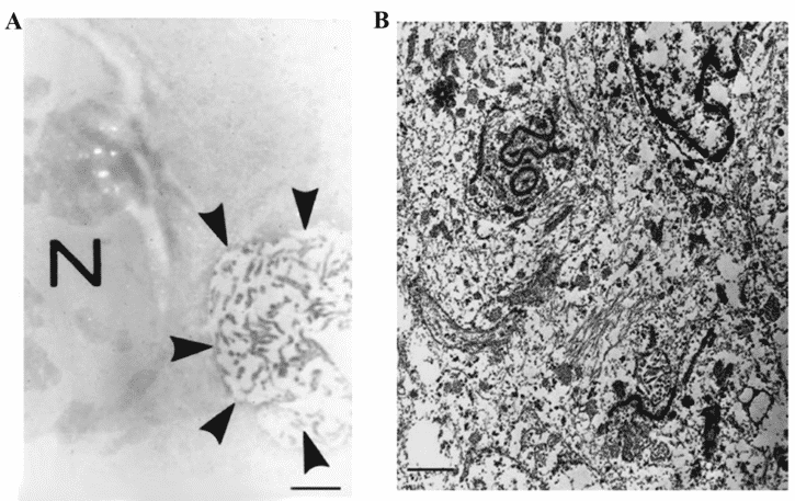 Figure 1. Diagnosis of ependymoma.
Over the years Creative Biostructure has been devoted to developing in-house EM platform and exploring cutting-edge technologies to use EM in diagnostic pathology. Based on our up-to-date EM platform, Creative Biostructure has gleaned extensive experience in various specimen preparations. Our team of seasoned scientists are ready to help the clients tackle some of the most challenging tasks.
Creative Biostructure promises to work closely with our customers to provide quality services of structural studies with unique biological insights.
Please feel free to contact us for a detailed quote.
Ordering Process

References
- Yuji Uematsu, The role of electron microscopy in the diagnosis of surgical pathology in the central nervous system. Med Mol Morphol. 2006; 39: 127-35.
- Diagnostic electron microscopy (https://en.wikipedia.org/wiki/Diagnostic_electron_microscopy).