Atomic Force Microscopy (AFM) Service
Atomic Force Microscope (AFM) is used to study the surface structure and properties of substances by detecting the extremely weak interatomic interaction between the surface of the sample to be tested and a miniature force-sensitive element. It can detect the physical properties of various materials and samples, including the nanometer region's morphology in the atmosphere and liquid environment, or directly perform nanomanipulation. In addition to providing surface images, AFM can also provide quantitative measurements of morphology, such as height differences and other dimensions. AFM has a wide range of applications and plays a vital role in various fields of scientific research and industry. The main principle of AFM is to fix one end of a microcantilever that is extremely sensitive to the weak force, and the other end has a tiny needle tip, which is in light contact with the sample surface. Because there is a feeble repulsive force between the atoms at the tip of the needle and the atoms on the surface of the sample, by controlling the constant force during scanning, The micro-cantilever with the tip will fluctuate in the direction perpendicular to the surface of the sample corresponding to the equipotential surface of the force between the tip and the surface atoms of the sample. Using the optical detection method or the tunnel current detection method, the position change of the micro-cantilever corresponding to each scanning point can be measured so that the information on the surface topography of the sample can be obtained.
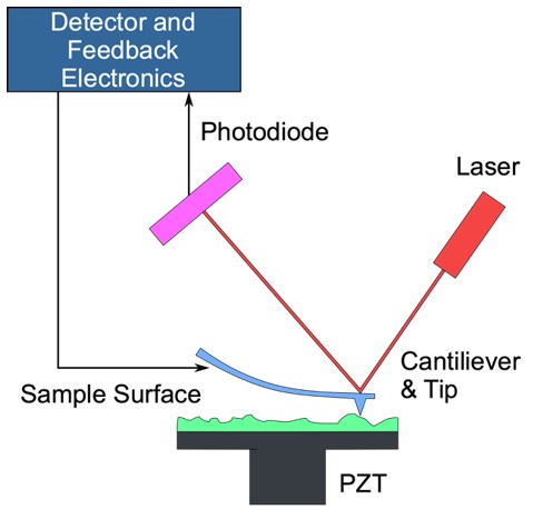 Figure 1.
Working principle of AFM.
Figure 1.
Working principle of AFM.
Imaging Modes of the Atomic Force Microscope
Contact Mode
Contact mode AFM is a repulsive mode. The probe tip makes soft "actual contact" with the sample. When the tip gently sweeps the surface of the sample, the force of the contact causes the cantilever to bend, and then the surface pattern of the sample is obtained. Normally, contact mode can produce stable, high-resolution images. Still, it is unsuitable for studying biological macromolecules, samples with low elastic modulus, and samples that are easy to move and deform.
Non-Contact Mode
In the non-contact mode AFM, the needle tip vibrates above the sample surface and never touches the sample, and the detector detects long-range forces such as van der Waals force and electrostatic force that do not damage the imaging sample. Due to the non-contact state, it is better to study soft or elastic samples. This mode is relatively difficult to manipulate, is generally unsuitable for imaging in liquids, and has few applications in biology.
Tapping Mode
Tapping AFM means that the microcantilever is forced to vibrate near its resonant frequency, and the oscillating tip gently taps the surface to contact the sample intermittently. There is little damage to the sample, and it is suitable for soft, brittle, and highly adhesive samples without damaging them. However, the disadvantage is that the scanning speed is slower than the contact modes.
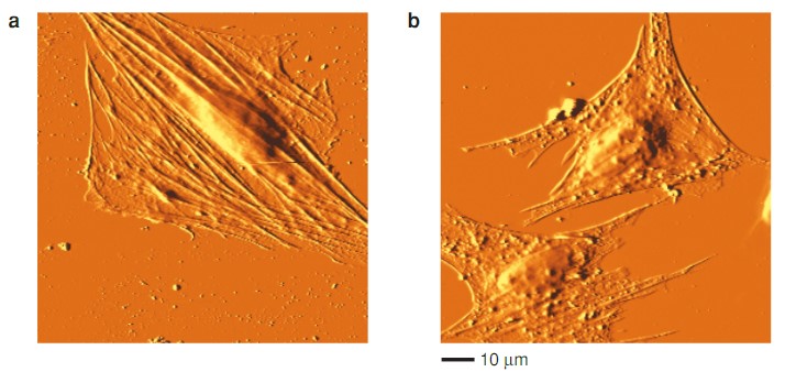 Figure 2. Examples of results of scanning cells with an AFM in contact mode in liquid.
Applications of Our Atomic Force Microscope Service
-
Application of AFM in Biology
- Observation of biological cell surface morphology
- Research on the structure and properties of biological macromolecules
- Observation of force spectrum curves between biological molecules
-
Application of AFM in Materials Science
- Topography observation
- Composition analysis
- Nanomaterials and powder material analysis
- Crystal growth observation
-
Application of AFM in Thin Film Technology
- Observation and determination of membrane surface structure
- Observation of membrane surface morphology
- Study on fouling degree of membrane surface
- Film formation mechanism research
-
Application of AFM in Physical Science
- Surface topography
- surface reconstruction
- Surface electronic states and dynamic processes
- Charge density
Creative Biostructure's scientists can offer you highly accurate measurements such as surface topography, dopant distribution, magnetic domain features, and various other sample properties to give you the information you need to do great work. Please feel free to contact us for more information or a detailed quote.
Ordering Process

References
- Lang K M., et al. Conducting atomic force microscopy for nanoscale tunnel barrier characterization. Review of Scientific Instruments. 2004, 75(8): 2726-2731.
- Binnig G, Quate C F, Gerber C. Atomic force microscope. Physical Review Letters. 1986, 56(9): 930.
- Geisse N A. AFM and combined optical techniques. Materials Today. 2009, 12(7-8): 40-45.
- Horton M A., et al. New technologies in scanning probe microscopy for studying molecular interactions in cells. Expert Reviews in Molecular Medicine. 2000, 2(2): 1-19.


