Cryo-Electron Tomography (Cryo-ET)
Creative Biostructure offers services based on cryo-electron tomography (Cryo-ET) technology, a powerful tool in the fields of structural biology and biophysics. Our cryo-ET services provide molecular-resolution 3D images of unperturbed cellular landscapes, enabling in situ visualization of cellular molecular architecture. This allows us to gain insights into the dynamic interactions of these molecules within their native environment, ultimately leading to a comprehensive understanding of cellular processes.
What Is Cryo-Electron Tomography (Cryo-ET)?
Beyond the individual molecular components, understanding the context and interactions of these components is the true key to unlocking the secrets of cellular function. That's where cryo-ET takes center stage. When cells are taken apart or molecules are extracted from their native environment, valuable information about their native interactions is lost forever. This cutting-edge technology allows us to visualize cellular molecular structures right within their original context, without compromising their structural integrity.
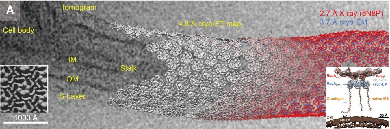 Figure 1. A tomographic slice of a C. crescentus cell showing the inner membrane (IM), outer membrane (OM), and surface layers (S-layer). (Turk
M, et al., 2020)
Figure 1. A tomographic slice of a C. crescentus cell showing the inner membrane (IM), outer membrane (OM), and surface layers (S-layer). (Turk
M, et al., 2020)
Cryo-ET utilizes an electron microscope to capture a series of 2D projection images acquired by tilting a biological sample held at cryogenic temperature. Through computational methods, the 3D images can be aligned and combined mathematically to generate a 3D (tomographic) reconstruction of the sample. A more comprehensive understanding of particle morphology is accessible by extracting individual slices in various orientations. Additionally, the 3D volume can be examined to resolve any ambiguities of the 2D images.
Our Workflow for Cryo-ET
Our cryo-ET services include a rigorous and systematic procedure to ensure the highest quality results. During the initial phase, we work closely with our clients to carefully prepare samples for vitrification and thinning, with a focus on preserving cellular structures in their unaltered, native state. Subsequently, the samples undergo observation and tomogram acquisition using an electron microscope. The acquired data is then subjected to tomogram reconstruction and further analysis.
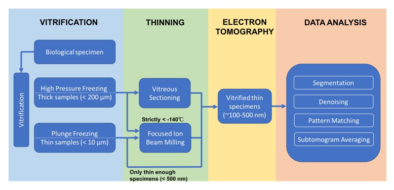 Figure 2. Schematic representation of the cryo-ET workflow.
What Can We Offer?
- In situ structural studies can achieve resolutions of 3-4 nm, and applying sub-tomogram averaging methods allows us to attain sub-nanometer (2-5 Å) resolution.
- We can apply cryo-ET to study a wide range of objects, from isolated proteins and organelles to small bacteria, viruses, or minicells. By incorporating the focused-ion beam (FIB)-milling method, we can extend our sample range to eukaryotic cells, multicellular organisms, and tissues.
- Correlation photoelectron microscopy (CLEM) aids in identifying and precisely localizing features in cellular landscapes. Cryo-super-resolution optical wave imaging (cryo-SOFI) enhances the accuracy of localizing fluorescently labeled proteins beyond the diffraction limit.
- For specific research objectives, this technique can be integrated with various technologies, such as mass spectrometry, offering a comprehensive approach to structural and functional analysis.
- Our cryo-ET service can also effectively characterize drug delivery vehicles, providing information on their structure, cargo, and interactions with tissues and cells.
- Versatile data analysis, including 2D suitability and averaging, and 3D reconstruction from existing data sets, and 3D structure refinement.
Highlights of Our Cryo-ET Service
- Cutting-Edge Technology: We utilize the latest advancements in cryo-ET to provide high-resolution 3D imaging of cellular structures, ensuring you get the most accurate insights.
- Expert Team: Our team of experienced scientists and researchers are dedicated to delivering precise and reliable results. We understand the unique challenges of each project and tailor our approach accordingly.
- Customized Solutions: We work closely with our clients to develop personalized strategies for sample preparation and analysis, ensuring that each project's goals are met effectively.
- Comprehensive Insights: Our service doesn't just stop at imaging. We assist you in interpreting the data, offering a holistic understanding of the structural and functional aspects of your samples.
As experts in the field of structural biology, Creative Biostructure has many years of experience in macromolecular structure research. Our specialty lies in offering cutting-edge cryo-ET solutions for the comprehensive analysis of diverse samples, shedding light on structural characterizations and intricate interactions. We are unwavering in our commitment to delivering top-tier services, ensuring your research objectives are met with precision and excellence. Contact us for a detailed quote if you're interested in our cryo-ET services.
Ordering Process

References
- Lučić V, Rigort A, Baumeister W. Cryo-electron tomography: the challenge of doing structural biology in situ. Journal of Cell Biology. 2013, 202(3): 407-419.
- Turk M, Baumeister W. The promise and the challenges of cryo-electron tomography. FEBS Letters. 2020, 594(20): 3243-3261.

