Immunoelectron Microscopy Service
With over a decade of experience, Creative Biostructure is dedicated to help our clients localize molecules at the ultrastructural level by labeling them with specific antibodies based on our unparalleled cryo-EM platform. To perform Electron Microscopy (EM) service is an elaborate process that involves careful planning, meticulous execution, and intensive trouble-shooting. An effective combination of the most suitable components will greatly increase the chance for a successful EM project. With this concept in mind, Creative Biostructure has perfected our cryo-EM development services, and develop our immunoelectron microscopy services allowing the client to identify antibody/antigen complexes that localize to a particular subcellular organelle or compartment.
Immuno-electron microscopy is used to localize molecules at the ultrastructural level by labeling them with specific antibodies. The antibodies are visualized by electron-opaque markers (colloidal gold particles) attached to them. The effect is to produce an electron-dense label at the site of the antigen-antibody reaction. When the antigen is located inside of the cell, then transmission electron microscopy is required to see it. The labeling can be done pre-embedding or post-embedding (on thin sections).
When the antigen in question is located on the surface of the specimen, scanning electron microscopy can be used to see it. The electron dense label can then be viewed using the back scatter image on an appropriately equipped microscope.
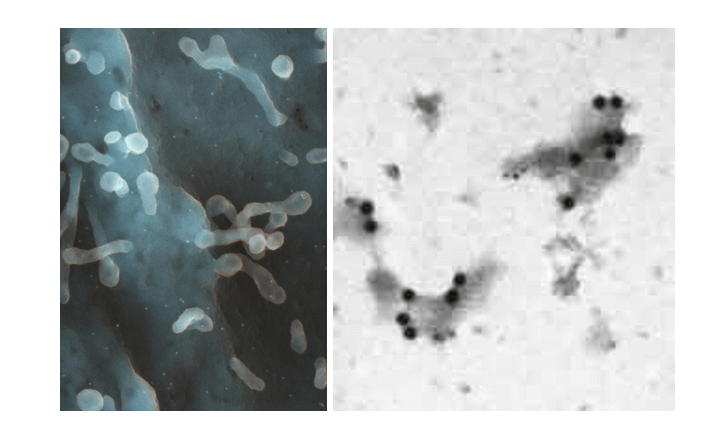 Figure 1. Immuno labeled spiny dendrite cell-SEM (left) and Double labeled cell membrane fragments (right).
This technique uses antibodies to detect the intracellular location of structures of particular proteins by electron microscopy. Ultra thin sections are labeled with antibodies against the required antigen and then labeled with gold particles. Gold particles of different diameters enable two or more proteins to be studied. By using a pre-embedding streptavidin-biotin technique with diaminobenzidine (DAB) as a chromagen which is silver enhanced, the tissue is post-fixed in osmium tetroxide, dehydrated and embedded in EPON/Araldite resin.
For example, paraffin embedded tissue (same tissue) sections are cut and screened by light microscopy using a streptavidin-biotin technique to determine the appropriate antibody concentration to be used for the EM immunogold procedure. Ultrathin sections from the same block(s) are cut, incubated with primary antibody and then later incubated with Protein A gold particles (size range is 5 nm to 20 nm). The gold particles bind to the Fc portion of the antibody and are detected by EM. A variation of the large block "pop-off" technique for immunoelectron microscopy is also available.
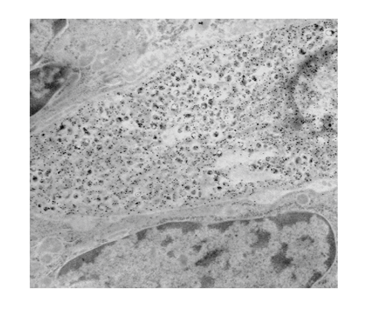 Figure 2. Protein A gold labeling of a prostatic endocrine-paracrine cell demonstrating localization of calcitonin (inset) to the neuroscretory granules.
We are more than happy to share our experience and help our customers with this tragically important step in immunoelectron microscopy and cryo-EM development. We like to be in touch with our customers so please feel free to contact us for a formal quote.
Ordering Process

Reference
- de Mesy Jensen et al. Large block embedding and "pop-off" technique for immunoelectron microscopy with applications to prostatic endocrine-paracrine cells. Ultrastruc. Pathol. 16:51, 1992.


