Exosome 3D Skin Model Assays
In the rapidly advancing fields of dermatology, regenerative skincare, and therapeutic aesthetics, proving the efficacy and safety of novel ingredients is paramount. Topical exosomes are emerging as a powerhouse active ingredient for skin applications, but traditional 2D cell culture assays fail to replicate the complex, multi-layered environment of human skin.
Our Exosome 3D Skin Model Assays bridge this gap. This platform is designed to provide comprehensive data on both the efficacy (e.g., anti-aging, wound healing) and safety (e.g., irritation, cytotoxicity) of your exosome active ingredient or finished formulation. We use reconstructed human skin models to quantitatively measure the biological effects of your candidate, providing the hard data needed to validate its potential for exosomes for skin rejuvenation.
Why Use 3D Skin Models for Exosome Testing?
Standard 2D cell assays lack a functional stratum corneum, the skin's primary barrier, making them unsuitable for testing topical exosomes. 3D models offer a superior, more clinically relevant alternative for both R&D and product validation.
- Physiological Relevance: Our models feature a fully differentiated epidermis and dermis, mimicking real human skin structure.
- Realistic Application: Allows for the topical application of both raw materials and finished products (e.g., exosomes skin cream), mirroring real-world use.
- Efficacy and Safety Assessment: Provides a robust platform for evaluating both desired outcomes (e.g., collagen synthesis) and potential adverse effects (e.g., skin irritation).
- Complex Endpoint Analysis: Enables advanced readouts not possible in 2D culture, such as changes in tissue architecture and multi-layer protein expression.
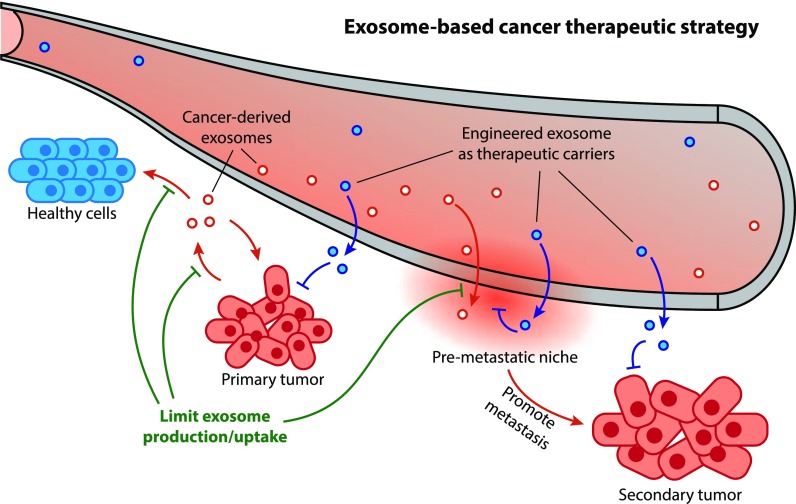
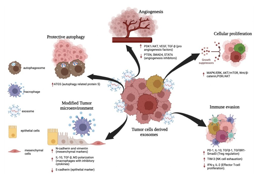
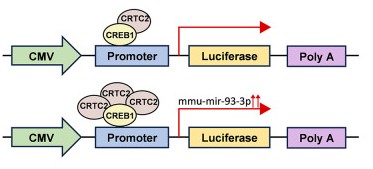
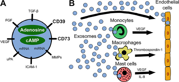
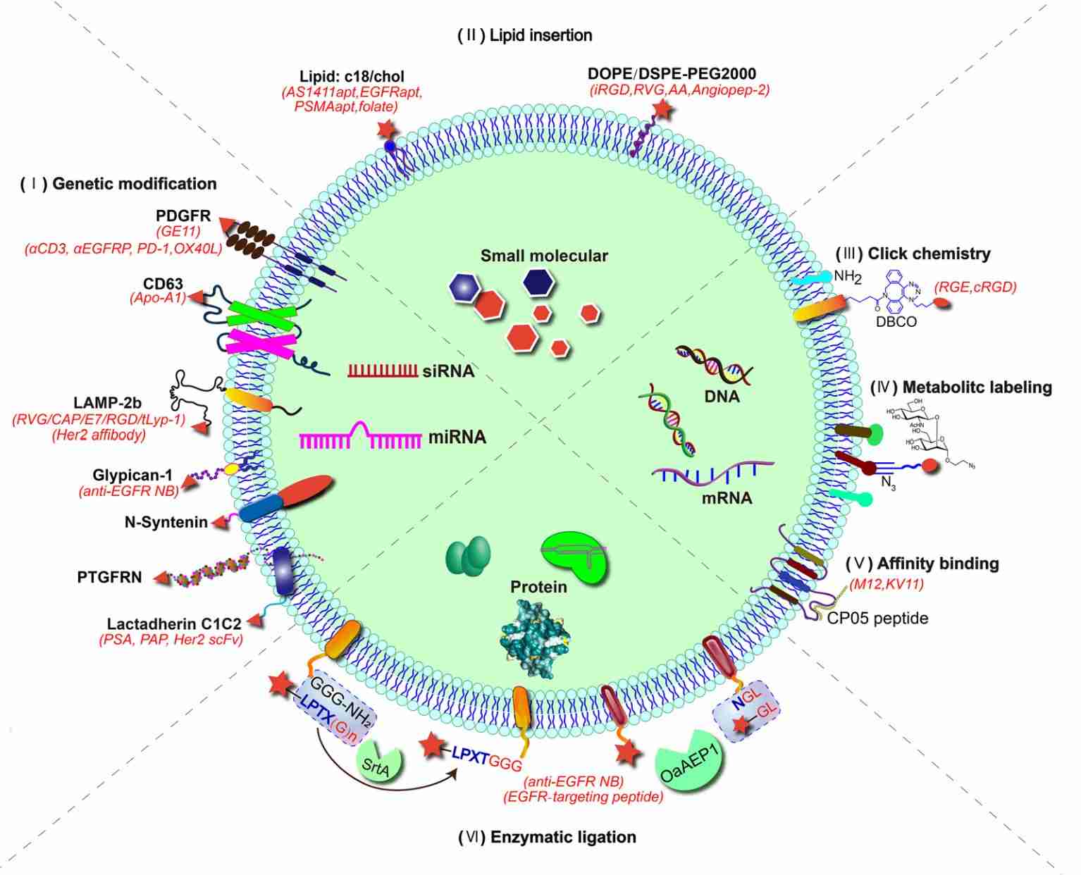
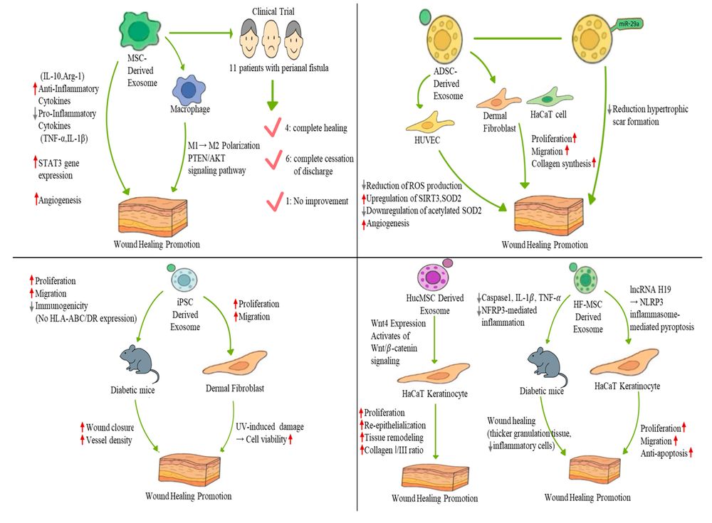 Figure 1. Representative mechanisms of stem cell-derived exosomes in cutaneous wound healing. (Jo C, et al., 2025)
Our Platform of 3D Skin Model Assays
Our comprehensive portfolio of assays is structured to provide the scientific proof needed to support your key research goals or product marketing claims, covering everything from rejuvenation to safety.
- For "Anti-Aging, Rejuvenation & Skin Tightening" Claims
Validate the core benefits of your regenerative skincare candidate. We measure the direct impact of your exosomes on the skin's structural integrity.
Key Endpoints Measured: Collagen I & III Synthesis, Elastin Expression, Epidermal Thickness, Dermal Density, Inhibition of MMP-1 (collagen-degrading enzyme).
- For "Wound Healing & Barrier Repair" Claims
Provide proof for candidates aimed at skin repair and protection. We create a wound in the 3D model and test your exosome's ability to accelerate healing.
Key Endpoints Measured: Re-epithelialization Rate (Wound Closure Speed), Barrier Function Restoration (via TEWL), Structural Regeneration (via Histology).
- For "Anti-Inflammatory & Soothing" Claims
Perfect for validating candidates targeting sensitive or compromised skin. We induce inflammation (e.g., via UV or chemical stress) and measure your exosome's calming effect.
Key Endpoints Measured: Reduction of Pro-inflammatory Cytokines (e.g., IL-1α, IL-6, TNF-α), Reduction in Tissue Damage Markers.
- For "Skin Brightening & Pigmentation Control" Claims
For candidates targeting uneven skin tone. Using pigmented skin models, we can test your exosome's effect on melanin production.
Key Endpoints Measured: Melanin Content Quantification, Tyrosinase Activity Inhibition.
- For "Safety & Non-Irritating" Claims
Generate the essential safety data for your exosome active or formulation. All assays are performed according to internationally recognized guidelines.
Services Offered: Standardized assays for skin irritation, corrosion, and phototoxicity.
Advantages of Our 3D Model Platform
| Feature | Description | Your Benefit |
|---|---|---|
| Claim-Oriented Design | Our assays are structured to directly answer the questions needed to support your research goals or product marketing claims. | Get clear, actionable data that directly translates to your scientific story or product value. |
| Physiologically Relevant Data | We use validated, multi-layered reconstructed human skin, providing data that is far more predictive of clinical outcomes than simple 2D cell culture. | Increase your confidence in your candidate's performance before committing to expensive clinical trials. |
| Efficacy & Safety in One Platform | We offer a comprehensive solution, allowing you to test both the beneficial effects and the potential risks of your formulation in parallel. | Streamline your R&D process, saving time and resources by using a single expert provider for all your preclinical skin model testing. |
| Publication-Ready Quality | We provide high-resolution images and statistically analyzed quantitative data, delivered in a comprehensive report ready for regulatory submission or publication. | Accelerate your time-to-market and strengthen your scientific credibility with a robust, professional data package. |
Our 3D Skin Model Workflow
Our workflow is specifically designed for the complexities of 3D tissue culture and topical application testing, ensuring every step from application to analysis is robust and reproducible.
Key Stages of Our Service
Consultation and Model Selection
The process begins with a detailed consultation to select the precise 3D skin model for your objective—such as an epidermis, full-thickness, or pigmented model—and to define the key endpoints required to validate your research goals.
Model Equilibration and Pre-treatment
The 3D tissue models are received and allowed to equilibrate. For specific efficacy studies, such as anti-inflammation or anti-aging, a pre-treatment stressor like UV radiation may be applied to induce a measurable damage or stress response.
Topical Candidate Application
Your exosome candidate, whether as a raw material solution or a finished formulation like a cream, is carefully and realistically applied to the surface of the 3D skin model, mimicking real-world use.
Our 5-Step Assay Pathway
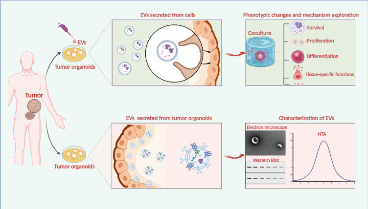
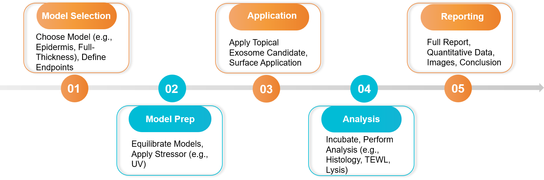 Figure 2. 3D Skin Model Assay Project Workflow. (Creative Biostructure)
Sample Requirements
- Exosome Test Article:
- Finished Formulations: Serums or creams are accepted. Please provide the formulation base (vehicle) as a negative control.
- Raw Material/Active Ingredient: Purified exosome solutions are accepted. Please provide the suspension buffer as a control.
- Controls: A vehicle/placebo control (the formulation without exosomes) is essential for a valid study.
Standard Deliverables
- A comprehensive project report with detailed methodology and protocols.
- Quantitative data for all key endpoints (e.g., collagen expression, cytokine levels, cell viability percentages).
- High-resolution, publication-quality images from histological analysis.
- A final consultation to discuss the data and its implications for your research or product claims.
Case Study
Case: Validating the Anti-Photoaging Effects of Adipose Stem Cell-Derived Exosomes on a 3D Skin Model
Background: Researchers hypothesized that exosomes from 3D-cultured adipose-derived stem cells (ADSC-Exos) could protect skin from UV-induced aging (photoaging). They needed a physiologically relevant model to prove this efficacy claim.
Methodology: A full-thickness 3D human skin model was used.
- Damage Induction: The skin models were exposed to UVB radiation to simulate sun damage.
- Topical Treatment: The irradiated models were then treated topically with the ADSC-Exo formulation.
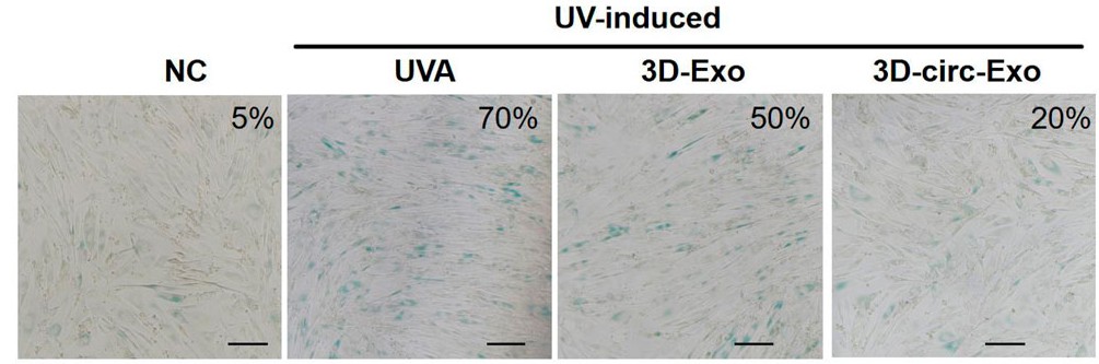 Figure 3. Histological cross-sections showed that the collagen density in the exosome treatment group was improved. (Zhang Y, et al., 2025)
- Endpoint Analysis: After incubation, the tissue was analyzed for key biomarkers of skin aging: collagen I expression, elastin expression, and the activity of the matrix-degrading enzyme MMP-1.
Key Findings:
- Compared to the UV-damaged control, the skin models treated with topical exosomes showed a significant increase in collagen I and elastin expression.
- The exosome treatment also significantly inhibited the activity of MMP-1, the enzyme responsible for breaking down collagen after sun exposure.
- Histological analysis confirmed a denser, more organized dermal structure in the exosome-treated group, indicative of a regenerative skincare response.
Conclusion: The 3D skin model assay provided definitive proof of efficacy. It validated that the ADSC-exosome formulation could not only protect the skin from UV damage but also actively promote skin repair by boosting key structural proteins.
Ready to validate topical exosomes in 3D skin models? We design targeted studies and produce quantitative efficacy and safety data. Contact us for a free consultation.
References
- Jo C, Choi YJ, Lee TJ. Therapeutic Potential of Stem Cell-Derived Exosomes in Skin Wound Healing. Biomimetics (Basel). 2025 Aug 20;10(8):546.
- Zhang Y, Zhou F, Nie G, et al. 3D-cultured hADSCs-derived exosomes deliver circ_0011129 to synergistically attenuate skin photoaging. Front Genet. 2025 Sep 3;16:1627472.
Frequently Asked Questions
For any inquiries, our support team is ready to help you get technical support for your research and maximize your experience with Creative Biostructure.