Exosomal Glycosylation Analysis Services
Exosomes, as pivotal messengers in intercellular communication, carry a diverse array of glycoproteins that serve as sophisticated molecular codes for cell recognition and immune regulation. Creative Biostructure offers a specialized exosomal glycosylation analysis service to illuminate this hidden layer of biological information. Leveraging advanced lectin microarrays and mass spectrometry technologies, our service provides precise mapping of glycan structures and glycosylation sites, empowering clients to decipher disease-specific glycocodes and accelerate biomarker discovery.
Why is Exosome Glycosylation Analysis Important?
Exosomal glycosylation analysis decodes the biological information embedded in the sugar coats (glycans) attached to exosomal surfaces. These glycans act as specialized "identity cards" and "navigation systems," directly determining how exosomes are recognized, captured, and internalized by recipient cells. This process fundamentally regulates intercellular communication and immune responses.
The profound significance of this analysis lies in three key areas:
- Uncovering Disease Mechanisms & Biomarkers
Profiling disease-specific "glycosignatures" on exosomes through liquid biopsies enables the discovery of highly sensitive biomarkers for applications including early diagnosis, patient stratification, and disease monitoring.
- Enabling Targeted Therapeutic Strategies
Understanding exosomal glycosylation opens the door to engineering next-generation delivery systems. By modifying surface glycans, we can design "smart" exosomes that precisely target specific tissues or cells, vastly improving the efficacy and reducing the side effects of therapeutic cargo delivery.
- Revealing Fundamental Biology
This analysis provides critical insights into the fundamental biological functions of exosomes themselves, helping to answer why certain exosomes are selectively taken up by particular cell types and how they ultimately influence cell fate.
In essence, analyzing exosomal glycosylation is not just about mapping sugar structures; it's about accessing a rich layer of functional information that drives both physiological processes and pathological states, holding immense potential for revolutionizing diagnostics and therapeutics.
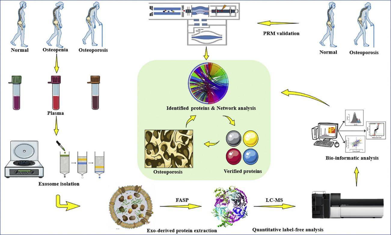
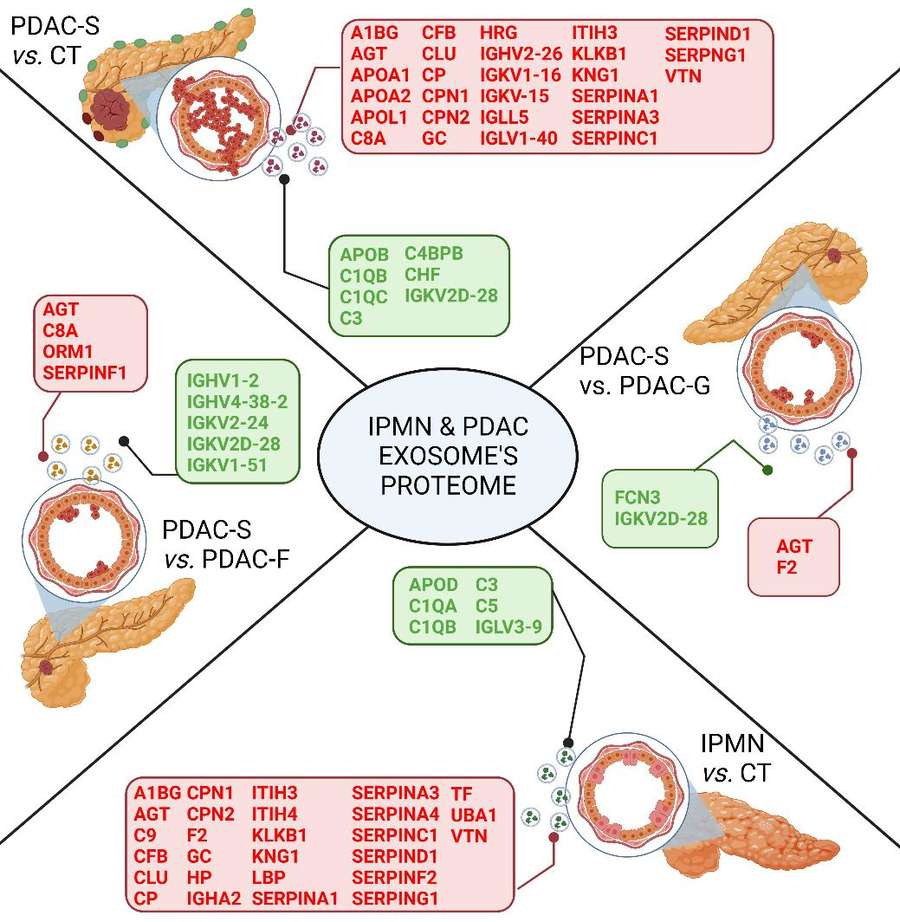
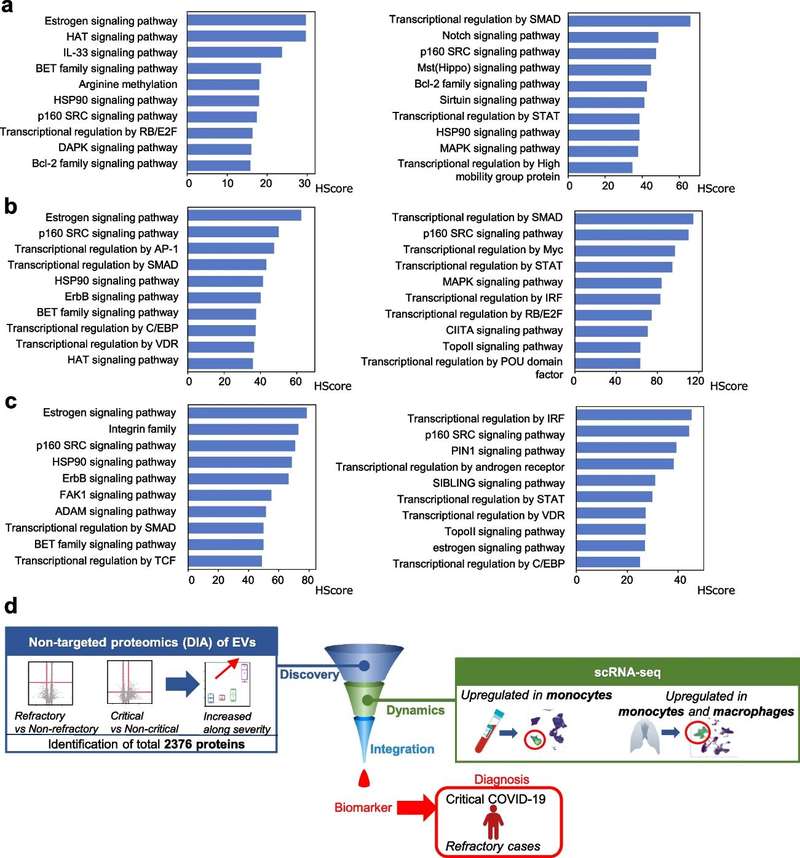
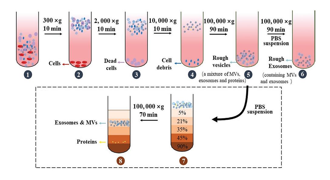
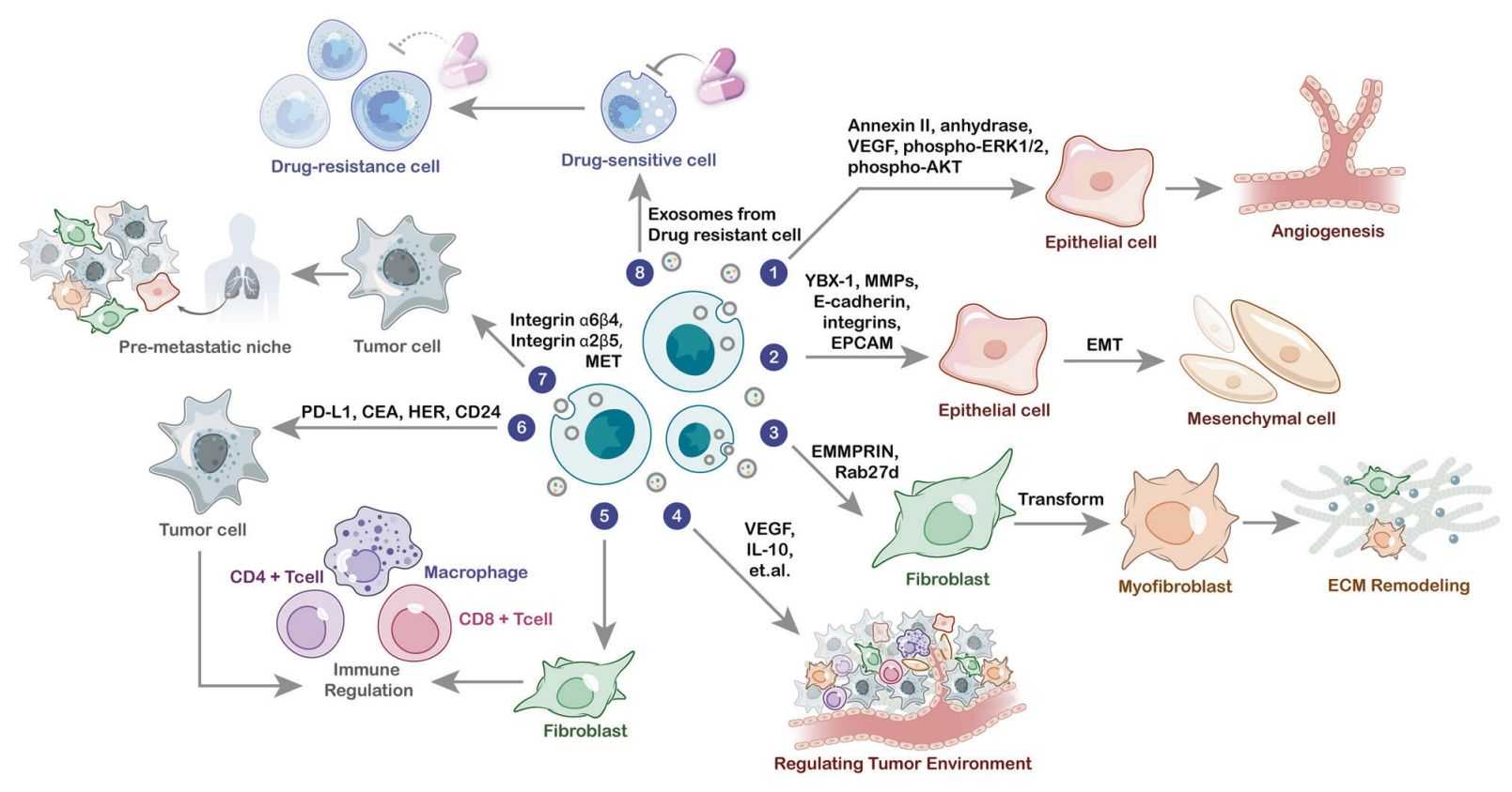
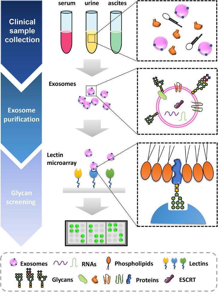 Figure 1. Glycosylation profile of exosomes can serve as biomarkers of disease states. (Lin S, et al., 2020)
Our Exosomal Glycosylation Analysis Service
Beginning with rigorous exosome isolation and purification from your provided samples (e.g., cell culture supernatant, blood plasma), we ensure the integrity of the native glycan structures. Our integrated technology platform then employs advanced techniques, including lectin microarray for high-throughput glycan profiling and high-sensitivity mass spectrometry for precise structural characterization and site-specific mapping of glycosylation.
The power of our service lies in the depth of our bioinformatics analysis. We deliver more than just raw data; we provide a biologically interpreted report that includes:
| Comprehensive Glycan Profiling | Identification and relative quantification of N-linked and O-linked glycans. |
| Glycosite Mapping | Precise localization of glycosylation sites on carrier proteins. |
| Comparative Analysis | Uncovering disease-specific or treatment-induced alterations in glycosylation patterns. |
| Functional Interpretation | Linking glycan signatures to potential biological functions and pathways. |
Workflow of Exosomal Glycosylation Analysis Service
Our service follows a rigorous, multi-stage workflow to ensure precise and comprehensive characterization of exosomal surface glycosylation:
Sample Receipt & Quality Control
We begin by accepting your submitted samples (e.g., cell culture supernatant, blood plasma, or serum). The received samples undergo immediate quality control, where we validate exosome presence and integrity using Nanoparticle Tracking Analysis (NTA) for concentration/size distribution and Western Blot for specific exosomal marker detection.
Exosome Isolation & Purification
We employ advanced isolation techniques tailored to your sample type, including ultracentrifugation, size-exclusion chromatography, or immunoaffinity capture, to ensure high-purity exosome recovery. The purified exosomes are subsequently validated for purity and protein content prior to glycan analysis.
Glycan Release & Labeling
Glycans are enzymatically or chemically released from the exosomal surface proteins and lipids. The released glycans are then purified and fluorescently labeled, enabling highly sensitive detection and quantification in subsequent analytical steps.
Glycan Profiling & Structural Analysis
We employ an integrated analytical approach:
- Lectin Microarray: For high-throughput screening of glycan-binding patterns and initial glycan fingerprinting.
- Liquid Chromatography-Mass Spectrometry (LC-MS/MS): For in-depth structural characterization, including identification of glycan compositions, linkages, and branching patterns, as well as site-specific mapping of glycosylation.
Bioinformatics & Data Interpretation
The raw data undergoes advanced bioinformatics processing for:
- Glycan Structural Identification & Quantification
- Comparative Glycoprofiling to identify significant alterations between sample groups
- Functional Annotation linking glycan changes to biological pathways and potential biomarker associations
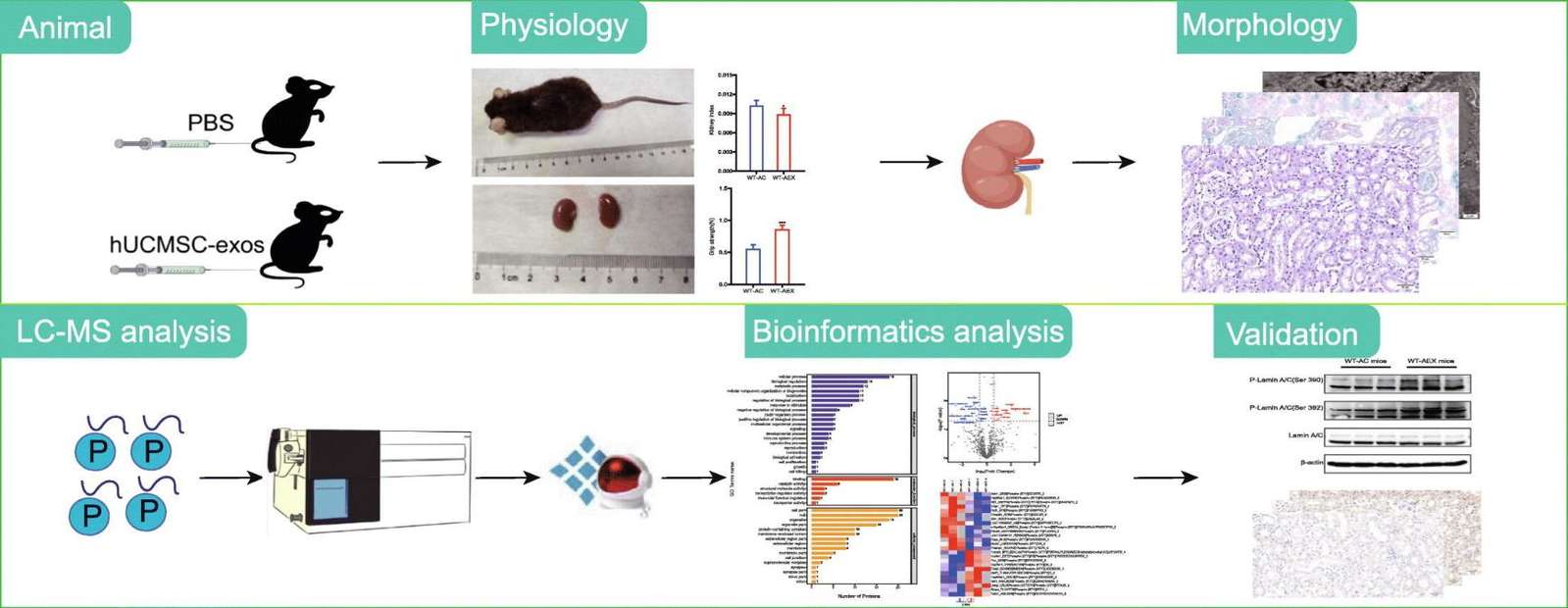
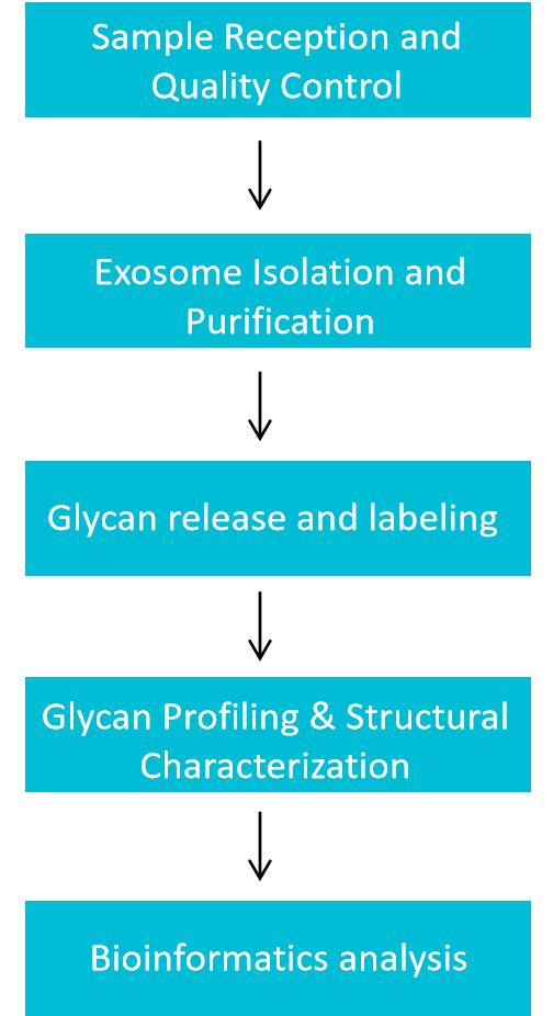 Figure 2. Exosomal glycosylation analysis service workflow. (Creative Biostructure)
Technical Advantages of Our Exosomal Glycosylation Analysis Service
- High Specificity: Distinguishes isomeric glycan structures (e.g., α2,3- vs. α2,6-sialylation) via high-resolution LC-MS/MS.
- Ultra-Sensitivity: Detects low-abundance glycosylation (down to pg level) to capture rare but biologically significant changes.
- Reproducibility: Standardized protocols and quality control (QC) samples (e.g., reference exosomes) ensure intra- and inter-batch CV < 10%.
- Customization: Supports personalized detection strategies (e.g., focused analysis of specific glycosylation types) and data delivery formats
Case Study
Case: Comprehensive Overview the Role of Glycosylation of Extracellular Vesicles in Cancers
Background
Colorectal cancer (CRC) represents a significant global health challenge and burden. This study profiles the aberrant glycosylation patterns on the surface of CRC cell-derived exosomes and proposes them as potential biomarkers for tumor characterization.
Methods
- The interactions of 27 lectins with exosomes from three CRC cell lines (SW480, SW620, HCT116) and one normal colon epithelial cell line (NCM460) have been analyzed by the lectin microarray.
- The expression of glycosylation related genes within cells has been analyzed by high-throughput quantitative polymerase chain reaction (HT-qPCR).
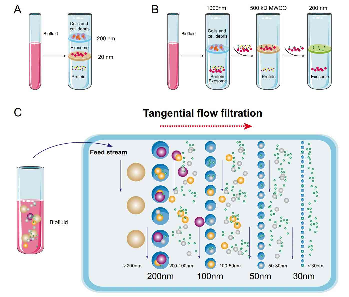
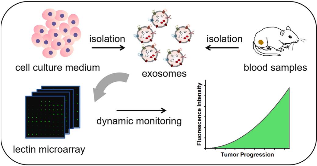 Figure 3. Glycan profiling of exosomes based on lectin microarrays. (Sun X, et al., 2024)
Conclusion
The results reveal that glycan expression pattern of exosome is linked to specific CRC subtypes, and regulated by glycosyltransferase and glycosidase genes of mother cells. The findings illuminate the potential of glycosylation molecules on the surface of exosomes as reliable biomarkers for diagnosis of tumor at early stage and monitoring of cancer progression.
Creative Biostructure leverages its integrated technology platform and specialized analytical team to provide clients with an end-to-end solution spanning from glycan structural analysis to functional interpretation. Whether exploring disease mechanisms, discovering novel biomarkers, or developing targeted delivery systems, we deliver robust data support and technical assurance. Contact us to discuss and obtain detailed technical proposals and consulting services tailored to your specific research needs.
References
- Lin S, Zhou S, Yuan T. The "sugar‐coated bullets" of cancer: Tumor‐derived exosome surface glycosylation from basic knowledge to applications. Clinical and Translational Medicine. 2020, 10(6): e204.
- Sun X, Chen B, Shan Y, et al. Lectin microarray-based glycan profiling of exosomes for dynamic monitoring of colorectal cancer progression. Analytica Chimica Acta. 2024, 1316: 342819.
Frequently Asked Questions
For any inquiries, our support team is ready to help you get technical support for your research and maximize your experience with Creative Biostructure.