Stroke Exosome Research Services
Stroke research is a race against time and tissue loss. Addressing the complex cascade of Ischemia-Reperfusion Injury (IRI)—neuronal death, blood-brain barrier (BBB) breakdown, and neuroinflammation—requires a multifaceted approach. Exosomes are at the forefront of this battle. They serve as circulating biomarkers for prognosis and, crucially, as potent therapeutic vehicles (e.g., MSC-derived exosomes) that can cross the BBB to promote neurogenesis and angiogenesis in the injured brain.
We provide a holistic Stroke Exosome Research Solution. Unlike generic service providers, we offer a dedicated portfolio covering the entire translational spectrum. Whether you need to screen plasma biomarkers for post-stroke prognosis, validate the neuroprotective effects of stem cell exosomes in OGD models, or quantify infarct reduction in MCAO animals, our platform integrates molecular, cellular, and in vivo modules to accelerate your cerebrovascular research.
Critical Research Frontiers in Stroke
Stroke research targets the complex cascade of injury and the limited window for effective treatment.
- Ischemia-Reperfusion Injury (IRI): Restoring blood flow is necessary but causes secondary damage via oxidative stress. Understanding the intercellular signaling that exacerbates IRI is crucial for cytoprotection.
- The Neurovascular Unit (NVU): Stroke affects neurons, astrocytes, and endothelial cells simultaneously. Research focuses on the exosome-mediated crosstalk within the NVU that determines BBB integrity and tissue survival.
- Regenerative Limitations: The brain has limited self-repair capacity. Investigating how stem cell-derived exosomes can stimulate endogenous neurogenesis and angiogenesis offers a cell-free therapeutic alternative.
- Post-Stroke Inflammation: Long-term disability is often driven by chronic neuroinflammation. Modulation of microglial polarization (M1 vs M2) via exosomal signaling is a key therapeutic strategy.
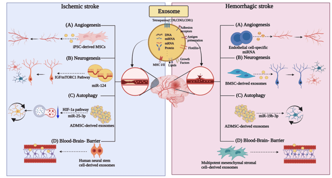 Figure 1. Exosome involvement in ischemic and hemorrhagic stroke through angiogenesis, neurogenesis, autophagy, and blood-brain barrier functions. (Lee EC, et al., 2022)
Comprehensive Service Portfolio for Stroke
We offer an integrated matrix of services tailored to your specific research focus, covering diagnosis, mechanism, and therapy.
| Research Phase | Our Specialized Approach & Solution | Key Services Applied |
|---|---|---|
| Biomarker Discovery (Diagnosis) | Circulating Profiling: We isolate exosomes from plasma or CSF and use Small RNA-Seq or Proteomics to find signatures of BBB damage or neuronal injury. | Exosomal Small RNA and miRNA Sequencing |
| Neuroprotection Mechanism (Cellular) | Ischemia Mimicry (OGD/R): We construct Oxygen-Glucose Deprivation/Reoxygenation (OGD/R) models in primary neurons. We test if exosomes rescue cells from apoptosis (TUNEL/Caspase-3). | Exosome Cellular Functional Assays |
| Therapeutic Development (Therapy) | Stem Cell Exosome Manufacturing: We produce high-quality MSC-derived exosomes (from Bone Marrow/Umbilical Cord) or NSC-Exos and verify their potency in promoting neurogenesis. | MSC and Stem Cell Derived Exosome Therapy |
| In Vivo Efficacy (Animal Models) | MCAO Model & Evaluation: We perform Middle Cerebral Artery Occlusion (MCAO) in rats/mice. We administer exosomes (IV/Intranasal) and assess infarct volume via TTC Staining. | In Vivo Exosome Functional Assays |
Core Technologies for Cerebrovascular Research
We highlight specialized technologies that are critical for validating stroke studies, ensuring high-impact data.
Oxygen-Glucose Deprivation (OGD/R) Model
The "In Vitro Stroke" Standard: To mimic the ischemic penumbra in a dish, simple hypoxia is not enough. We utilize specialized OGD/R systems where neurons are deprived of both oxygen and nutrients, followed by reperfusion. This allows for the precise screening of exosomal cargos (e.g., miRNAs) that confer resistance to oxidative stress and excitotoxicity before moving to animal studies.
Angiogenesis Tube Formation Assay
Restoring Blood Supply: Post-stroke recovery depends on re-vascularization. We employ HUVEC Tube Formation Assays on Matrigel to test the angiogenic potency of exosomes. We quantify total tube length and branching points to prove that your exosomes (e.g., from endothelial progenitor cells) can actively stimulate capillary network formation.
TTC Staining & Infarct Volumetry
Quantifying Brain Damage: The gold standard for assessing neuroprotection in vivo is measuring the infarct size. We utilize 2,3,5-Triphenyltetrazolium chloride (TTC) staining on brain sections from MCAO mice. This metabolic stain differentiates viable tissue (Red) from necrotic infarcts (White), providing a rigorous, quantitative readout of your exosome therapy's efficacy.
Application Spotlight: Engineered Exosomes Block Ferroptosis via Microglia
This analysis highlights the power of Surface Engineering to direct exosomes to specific immune cells, modulating the neuroinflammatory microenvironment to treat stroke.
Featured Technologies:
- Exosome Surface Functionalization (Targeting)
- Immunomodulation & Inflammation Assays
Literature Interpretation:
Ischemic stroke triggers a cascade of Ferroptosis (iron-dependent cell death) and pro-inflammatory microglial activation. To combat this, researchers developed an advanced Engineered Exosome system (M2pep-ADSC-Exo). These exosomes were surface-modified with M2pep, a peptide that specifically targets M2-type microglia. Upon administration in stroke models, the exosomes successfully crossed the BBB and accumulated in microglia, where they reduced susceptibility to ferroptosis via the Fxr2/Atf3/Slc7a11 axis. This targeted intervention not only prevented microglial death but also promoted a phenotypic shift toward the reparative M2 state, significantly suppressing neuroinflammation and improving neurological outcomes.
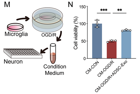 Figure 2. Schematic of conditioned medium experiment and CCK-8 assay results for N2a neuron survival rate after treatment with microglia-conditioned medium and ADSC-Exo. (Wang Y, et al., 2024)
Start Your Stroke Research Project
Leverage our comprehensive platform to accelerate your diagnostic or therapeutic program.
How It Works: Our Project Pathway
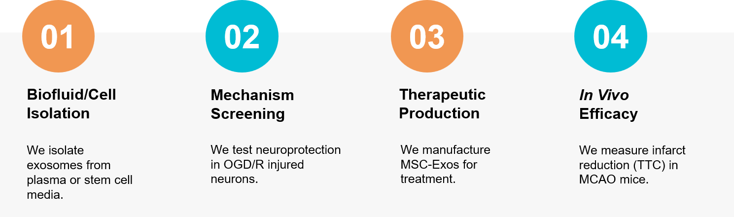 Figure 3. Integrated workflow for discovering stroke biomarkers, validating neuroprotection in OGD models, and assessing therapeutic efficacy via TTC staining in MCAO mice. (Creative Biostructure)
Ready to advance your stroke recovery research? Our neuro-vascular experts are available to build a custom study plan tailored to your needs. Contact us today to discuss your project.
References
- Lee EC, Ha TW, Lee DH L, et al. Utility of Exosomes in Ischemic and Hemorrhagic Stroke Diagnosis and Treatment. Int J Mol Sci. 2022 Jul 28;23(15):8367.
- Wang Y, Liu Z, Li L, et al. Anti-ferroptosis exosomes engineered for targeting M2 microglia to improve neurological function in ischemic stroke. J Nanobiotechnology. 2024 May 27;22(1):291.