TIRF Microscopy-Based Exosome Characterization Service
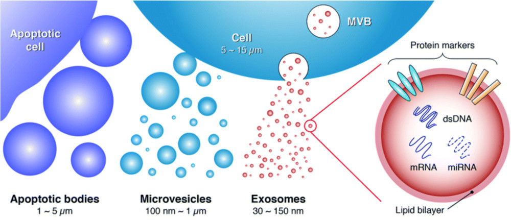
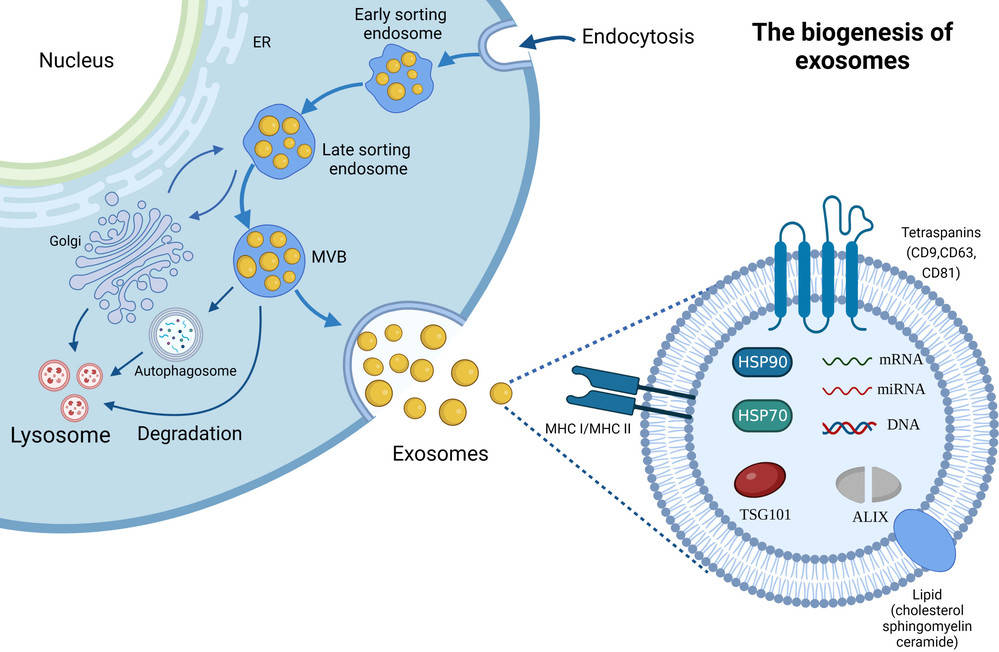
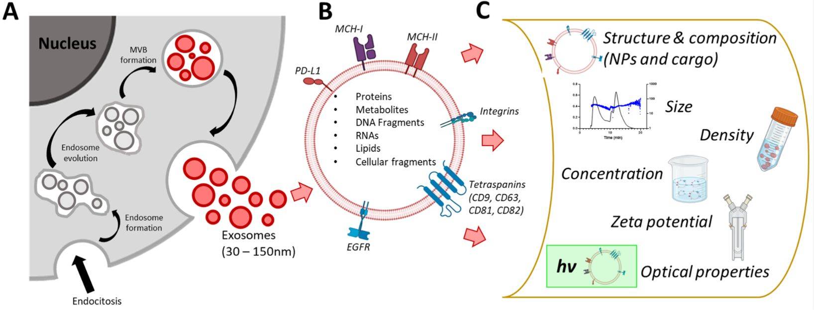
Exosomes are nanoscale extracellular vesicles that transport proteins, nucleic acids, and lipids between cells. They are essential mediators of intercellular communication and hold great promise as biomarkers and therapeutic tools. Traditional bulk methods such as Western blot or ELISA average signals across millions of vesicles, masking their true heterogeneity.
Total Internal Reflection Fluorescence (TIRF) Microscopy provides a powerful solution. By selectively exciting fluorophores within a shallow region at the glass interface, TIRF achieves an exceptional signal-to-noise ratio. This allows researchers to visualize and quantify the molecular composition of individual exosomes with precision. At Creative Biostructure, our TIRF service is carried out in compliance with MISEV2023 guidelines, ensuring reproducibility and transparency.
Why Choose TIRF for Exosome Analysis?
TIRF microscopy offers several unique benefits for exosome characterization:
- Exceptional signal-to-noise ratio by eliminating background fluorescence.
- Single-molecule sensitivity, enabling detection of low-abundance surface proteins such as CD9, CD63, and CD81.
- Quantitative stoichiometry that reveals the copy number of biomolecules per vesicle.
- High-throughput phenotyping of thousands of individual vesicles in a single run.
- Subpopulation identification and rare event detection within heterogeneous EV samples.
These advantages make TIRF an indispensable tool for biomarker discovery, therapeutic development, and quality control.
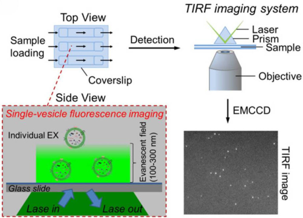 Figure 1. Single-Vesicle Fluorescence Imaging of Exosomes by TIRF Microscopy. (He D, et al., 2019)
TIRF vs. Other Exosome Characterization Methods
| Method | Analysis Level | Primary Information | Key Advantage | Limitation |
|---|---|---|---|---|
| TIRF Microscopy | Single-Vesicle | Molecular composition and stoichiometry | Unmatched sensitivity and quantitative accuracy | Requires immobilization |
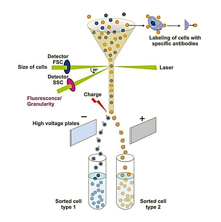
| Flow Cytometry | Single-Vesicle (specialized) | Size and co-localized fluorescence | High-throughput sorting | Limited sensitivity for small EVs |
| Nanoparticle Tracking Analysis | Particle Population | Size and concentration | Rapid sizing and quantification | No molecular information |
| Western Blot | Bulk Population | Average protein levels | Widely accessible | Masks heterogeneity and is non-quantitative |
TIRF provides complementary insights that cannot be obtained by population-based methods alone.
Our Comprehensive TIRF Imaging Assay Service
We provide an end-to-end solution, from consultation to data delivery, tailored to your project needs.
Our TIRF Service Capabilities
- Multi-color surface marker phenotyping to quantify exosome markers such as CD9, CD63, and CD81.
- Intra-vesicular cargo analysis for proteins or nucleic acids after permeabilization.
- Subpopulation stratification based on distinct molecular signatures.
- Receptor-ligand interaction studies with immobilized antibodies or substrates.
Step-by-Step TIRF Microscopy Exosome Characterization Workflow
Project Consultation
Our Ph.D. scientists work with you to design a TIRF imaging assay tailored to your research goals, including optimal sample preparation and labeling strategies.
Sample Preparation and Quality Control
We handle your samples with care, preparing them for analysis and performing initial QC checks.
Vesicle Immobilization and Staining
We utilize optimized protocols to either non-specifically adhere vesicles (e.g., via poly-L-lysine) or specifically capture them using antibody-coated surfaces for targeted analysis.
TIRF Imaging and Calibration
Imaging performed on high-resolution systems, with strict documentation of laser power, exposure time, and magnification. Calibration with single-molecule standards ensures quantitative accuracy.
Data Analysis and Quantification
We use validated software to detect thousands of vesicles as diffraction-limited spots. The intensity of these spots is analyzed to quantify the number of fluorescent molecules on each vesicle.
Reporting and Data Delivery
You receive a detailed report containing high-resolution images, quantitative data (e.g., population distributions, molecule counts), a full description of the methodology, and expert interpretation of the results.
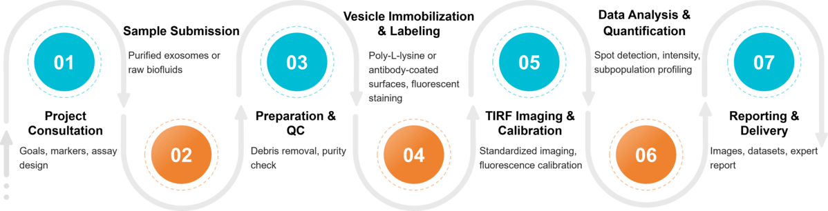 Figure 2. Project Workflow for Exosome Characterization by TIRF Microscopy. (Creative Biostructure)
Quality and Compliance Standards
Our service adheres strictly to MISEV2023 recommendations, including:
- Transparent reporting of microscope type, magnification, laser power, and exposure conditions.
- Calibration of the system for unbiased sensitivity assessment.
- Documentation of analysis software and parameters for reproducibility.
- Clear differentiation between vesicles and background artifacts.
Sample Requirements for TIRF Analysis
To guarantee reliable and reproducible outcomes, we recommend adhering to the sample submission guidelines outlined below.
| Requirement | Description |
|---|---|
| Sample Type | Pre-purified exosomes or raw material such as culture media, plasma, or urine |
| Purity | Free from apoptotic bodies, large debris, and excess protein aggregates |
| Concentration | ≥109 particles/mL for robust imaging |
| Volume | Minimum of 50-100 µL per run |
| Buffer Conditions | PBS or physiological buffer without detergents or high salt |
| Storage and Shipping | Fresh or frozen at -80 °C, shipped on dry ice |
What Deliverables Will You Receive
- Raw and processed TIRF microscopy images.
- Quantitative stoichiometry data for surface and intraluminal molecules.
- Subpopulation distribution and comparative analyses.
- Publication-ready figures, datasets, and methodological documentation.
- Technical report with expert interpretation.
Applications of TIRF in Exosome Research
Our service supports a broad range of research and development goals:
- Biomarker discovery by profiling disease-specific exosome markers.
- Therapeutic EV development with verification of surface modifications and drug loading.
- Fundamental biology through the study of vesicle heterogeneity and cell-to-cell communication.
- Quality control of engineered exosome preparations.
Why Choose Creative Biostructure?
- Cutting-edge TIRF platforms for high-sensitivity imaging.
- Expertise in both EV biology and advanced microscopy.
- Data fully compliant with MISEV2023 standards.
- Transparent and collaborative project management.
- Tailored reporting that supports academic publication and industrial R&D.
Case Study
Case: Single-Vesicle Profiling of Immune Cell-Derived Exosomes Using TIRF Microscopy
Objective: To characterize exosomes secreted by human and murine T lymphocytes at the single-vesicle level.
Methodology:
- Exosomes isolated by ultracentrifugation.
- About 8 × 10⁶ particles immobilized on antibody-coated coverslips.
- Labeled with exosomal markers CD9, CD63, and CD81.
- TIRF microscopy applied with a ~90 nm penetration depth to minimize background.
Results:
- Vesicles appeared as diffraction-limited fluorescent spots.
- Intensity analysis performed on thousands of vesicles.
- Identified distinct subpopulations and marker co-localization patterns.
Significance: TIRF microscopy revealed molecular diversity at the single-vesicle level, providing valuable insights for biomarker discovery and functional EV studies.
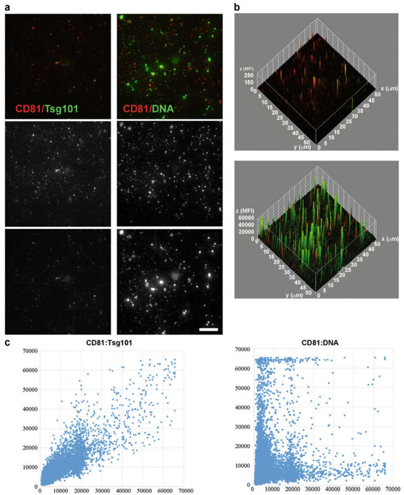 Figure 3. Exosome Analysis by TIRF Microscopy. (a) Representative TIRF images showing detected molecules in overlay and single-color formats (scale bar: 10 μm). (b) Surface plot graphs generated with ImageJ, illustrating spatial correspondence of fluorescence signals. (c) Correlation graphs between mean fluorescence intensity values obtained from TIRF images, confirming quantitative single-vesicle analysis. (Martín-Cófreces N B, et al., 2020)
At Creative Biostructure, we provide advanced TIRF microscopy services for precise exosome characterization. Our solutions support biomarker discovery, therapeutic EV development, and quality control. Contact us to discuss your project and receive a customized service plan.
References
- He D, Wang H, Ho S L, et al. Total internal reflection-based single-vesicle in situ quantitative and stoichiometric analysis of tumor-derived exosomal microRNAs for diagnosis and treatment monitoring. Theranostics. 2019, 9(15): 4494.
- Martín-Cófreces N B, Torralba D, Lozano-Prieto M, et al. TIRF microscopy as a tool to determine exosome composition. Stem Cell Renewal and Cell-cell Communication: Methods and Protocols. New York, NY: Springer US, 2020: 91-104.
- Welsh J A, Goberdhan D C I, O'Driscoll L, et al. Minimal information for studies of extracellular vesicles (MISEV2023): From basic to advanced approaches. Journal of Extracellular Vesicles. 2024, 13(2): e12404.
Frequently Asked Questions
For any inquiries, our support team is ready to help you get technical support for your research and maximize your experience with Creative Biostructure.