Exosome Morphology Analysis Service
At Creative Biostructure, we provide advanced exosome morphology analysis services for both research and industrial applications. Our expertise in high-resolution imaging allows accurate visualization and measurement of extracellular vesicles, helping clients evaluate vesicle integrity, purity, and structural features. This service is valuable for understanding the biological roles of exosomes, validating manufacturing processes, and ensuring compliance with international guidelines.
What Is Exosome Morphology Analysis and Why Is It Important
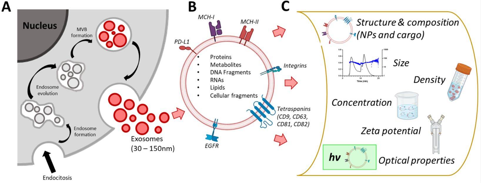
Exosome morphology analysis is the process of visualizing and measuring the size, shape, and surface features of extracellular vesicles (EVs) at the nanoscale. Exosomes are typically 30-150 nanometers in diameter, secreted by cells, and carry functional biomolecules such as proteins, nucleic acids, and lipids. Their morphology is a critical quality attribute that reflects biological function, sample integrity, and purity.
In research and industry, morphology analysis provides:
- Structural validation: Visual confirmation of vesicle integrity and detection of deformation or aggregation
- Purity assessment: Identification of co-isolated non-vesicular particles and potential contaminants
- Reproducibility assurance: Consistent morphology data for reliable experimental outcomes
- Application-specific insights: Linking vesicle structure to functional studies in diagnostics and therapeutics
Exosome morphology analysis is applied across multiple areas:
- Basic research: Studying vesicle biogenesis, heterogeneity, and intercellular communication
- Therapeutic development: Confirming the structural integrity of natural or engineered exosomes before clinical use
- Industrial production: Serving as a quality control checkpoint alongside nanoparticle tracking analysis and biochemical profiling
By combining visual evidence with quantitative measurements, exosome morphology analysis plays an essential role in advancing extracellular vesicle science and ensuring high standards for translational applications.
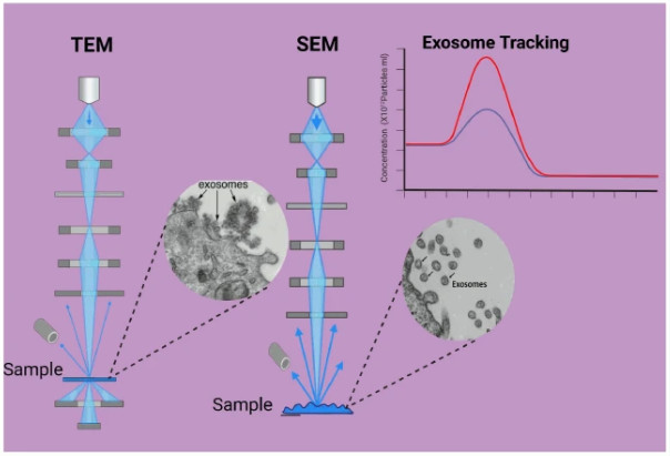 Figure 1. Schematic representation of exosome morphology analysis using transmission electron microscopy (TEM) and scanning electron microscopy (SEM). (Saleem M, et al., 2024)
Techniques We Offer for Exosome Morphology Analysis
We apply multiple complementary imaging methods to capture a complete picture of exosome structure:
| Technique | Key Purpose | Sample State | Resolution & Detail Level | Best Suited For | Advantages | Limitations |
|---|---|---|---|---|---|---|
| Transmission Electron Microscopy (TEM) | High-resolution structural imaging of exosome TEM samples | Fixed and stained (dehydrated) | Nanometer-scale; clear visualization of lipid bilayer | Vesicles of all sizes; morphology validation | Widely used; standardized protocols; excellent image clarity with contrast stains | Requires dehydration; may introduce artifacts if not optimized |
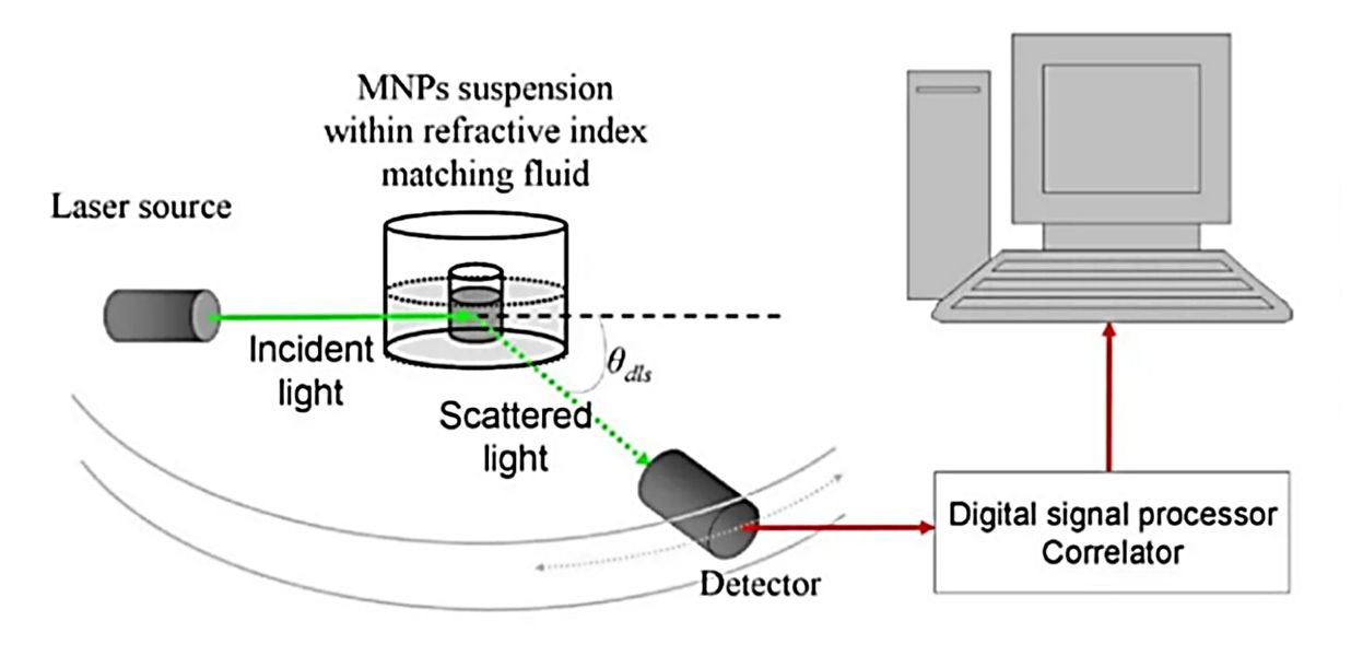
| Cryo-Electron Microscopy (Cryo-EM) | Native-state exosome ultrastructure imaging | Vitrified (hydrated) | Nanometer-scale; detailed bilayer and internal features | Quantitative analysis of all particles in a given volume | Preserves natural morphology; avoids dehydration artifacts; quantitative | More complex and costly; requires cryo-preparation |
| Scanning Electron Microscopy (SEM) | Surface topology imaging of exosomes under microscope | Fixed and coated | High surface detail; 3D-like appearance | Larger vesicles; identifying surface irregularities | Reveals surface texture; applicable to a wide size range | Limited internal detail; high magnification for smallest vesicles is harder to interpret |
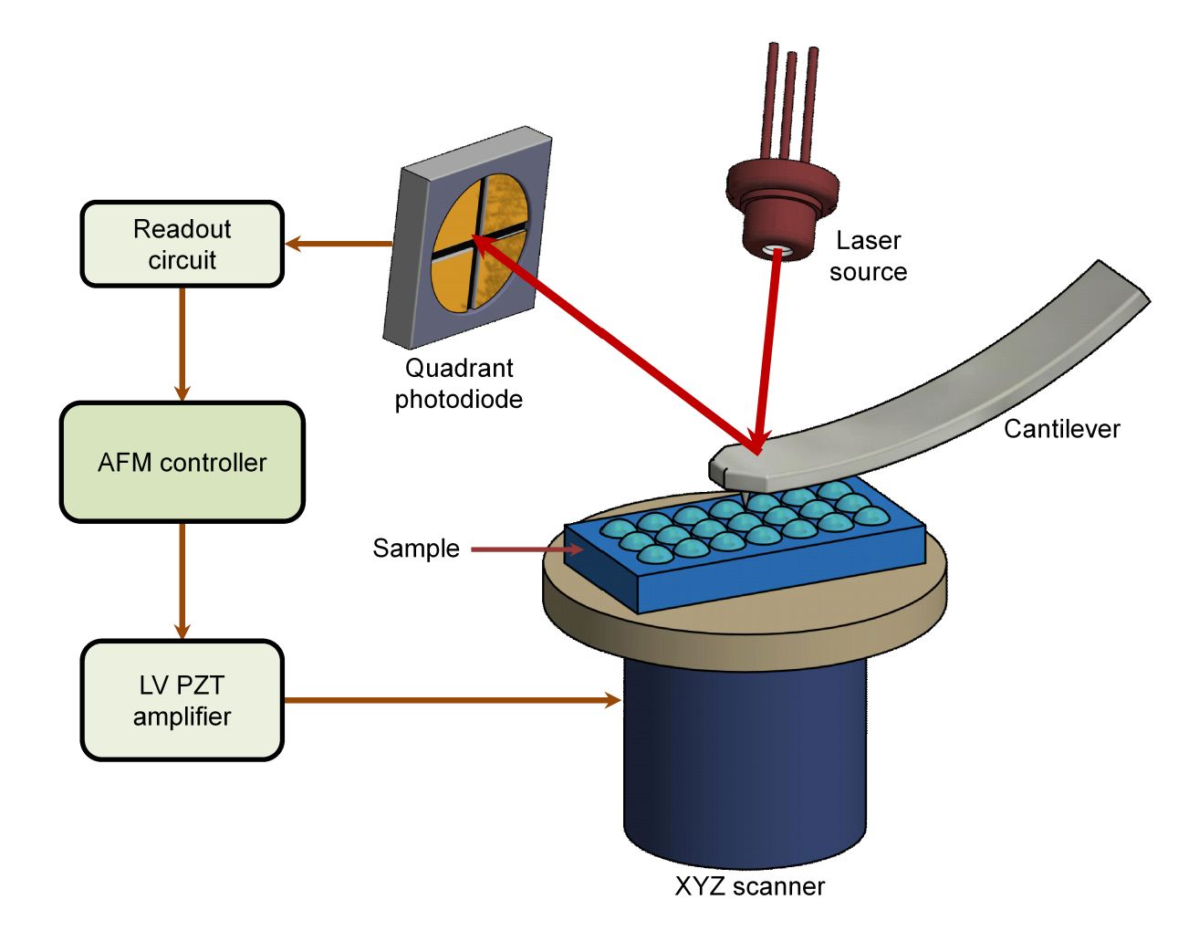
| Atomic Force Microscopy (AFM) | Nanomechanical and topographical mapping | Ambient or liquid | Nanometer-scale height and surface profile | Surface structure studies; vesicle stiffness measurements | Operates in liquid; measures physical properties; complements EM data | Smaller imaging area; slower scanning speed |
Our Compliance with International Guidelines
We follow MISEV recommendations and internal best practices to ensure that our imaging results are accurate, reproducible, and transparent:
- Detailed reporting of instrumentation, software, and acquisition settings.
- Documentation of sample preparation, including fixation, adsorption, and staining procedures.
- Provision of both high- and low-magnification images to capture vesicles in context and at full resolution.
- Use of appropriate controls to distinguish true vesicles from co-isolated particles.
Our Workflow for Exosome Morphology Analysis
Sample Receipt and Pre-Processing
Verification of sample quality and removal of large debris or unwanted particles.
Preparation for Imaging
Selection of hydrated or dehydrated protocols depending on the analytical technique.
High-Resolution Imaging
Capturing images using TEM, cryo-EM, SEM, or AFM.
Image Analysis
Measurement of vesicle size distribution, morphology, and structural integrity.
Data Delivery
Provision of a technical report, high-resolution image files, annotated image panels, and statistical data.
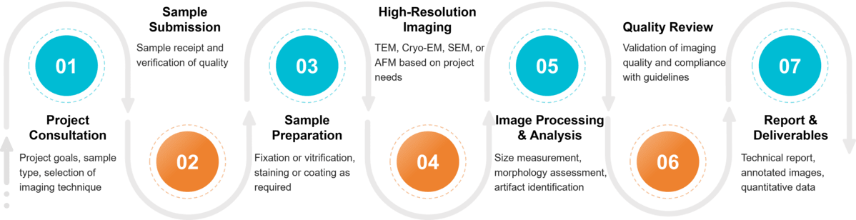 Figure 2. Project Workflow for Exosome Morphology Analysis. (Creative Biostructure)
Sample Requirements and Compatibility
| Sample Type | Minimum Volume | Preparation and Storage | Shipping Guidelines |
|---|---|---|---|
| Purified Exosomes | ≥ 100 µL | Resuspend in PBS or preferred buffer; store at -80 °C | Ship on dry ice |
| Plasma, Serum, Urine | ≥ 1 mL | Store at -80 °C immediately after collection | Ship on dry ice |
| Cell Culture Supernatants | ≥ 5 mL | Clarify by centrifugation to remove cells and debris; store at -80 °C | Ship on dry ice |
| Other Biological Fluids* | Variable | Follow matrix-specific preparation; contact us for details | Ship on dry ice |
* Examples include bile, breast milk, plant-derived fluids, cerebrospinal fluid (CSF), amniotic fluid, and tissue homogenates. Please inquire for tailored handling instructions.
What Deliverables Will You Receive
- High-resolution images showing exosome ultrastructure.
- Quantitative data for exosome measurement and morphology assessment.
- Annotated image panels highlighting structural features.
- A technical report describing methods, findings, and interpretations.
Why Choose Creative Biostructure
- Expertise: Years of hands-on experience in exosome characterization and exosome analytics.
- Advanced Equipment: Access to cutting-edge TEM, cryo-EM, SEM, and AFM systems.
- Quality Standards: Strict adherence to international guidelines to ensure reproducible, publication-ready data.
- Custom Solutions: Tailored packages for research, preclinical development, and industrial-scale production.
Case Study
Case: Morphological Characterization of hucMSC-Derived Exosomes in Cardioprotection Research
Researchers investigated the protective role of human umbilical cord mesenchymal stem cell-derived exosomes (hucMSC-exo) against hypoxia/reoxygenation (H/R)-induced pyroptosis in cardiomyocytes. The study identified miR-100-5p as a key exosomal component that suppresses FOXO3 expression, thereby inhibiting NLRP3 inflammasome activation, reducing IL-1β and IL-18 secretion, and protecting cells from inflammatory programmed cell death.
Creative Biostructure's Contribution
Creative Biostructure provided high-resolution TEM morphological characterization to validate vesicle integrity and size distribution. Using our MagHelix TEM platform, the study confirmed that isolated hucMSC-exo displayed uniform morphology with an average diameter of approximately 100 nm, consistent with the expected structural profile of extracellular vesicles. This morphological verification ensured that functional experiments were conducted on well-defined and intact exosome preparations, strengthening the study's conclusions regarding the link between vesicle structure and biological activity.
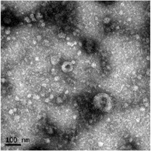 Figure 3. TEM image showing the morphology of purified exosomes derived from hucMSC-exo. The vesicles exhibit typical round or cup-shaped structures with a diameter of approximately 100 nm. Scale bar: 100 nm. (Liang C, et al., 2021)
At Creative Biostructure, we combine advanced imaging technologies with strict quality standards to deliver accurate and reproducible exosome morphology analysis results. Whether for basic research, therapeutic development, or industrial quality control, our team can provide the structural insights you need. Contact us to discuss your project and request a tailored service plan.
References
- Liang C, Liu Y, Xu H, et al. Exosomes of human umbilical cord MSCs protect against hypoxia/reoxygenation-induced pyroptosis of cardiomyocytes via the miRNA-100-5p/FOXO3/NLRP3 pathway. Frontiers in Bioengineering and Biotechnology. 2021, 8: 615850.
- Saleem M, Shahzad K A, Marryum M, et al. Exosome-based therapies for inflammatory disorders: a review of recent advances. Stem Cell Research & Therapy. 2024, 15(1): 477.
- Welsh J A, Goberdhan D C I, O'Driscoll L, et al. Minimal information for studies of extracellular vesicles (MISEV2023): From basic to advanced approaches. Journal of Extracellular Vesicles. 2024, 13(2): e12404.
Frequently Asked Questions
For any inquiries, our support team is ready to help you get technical support for your research and maximize your experience with Creative Biostructure.