Tunable Resistive Pulse Sensing (TRPS)-Based Exosome Characterization Service
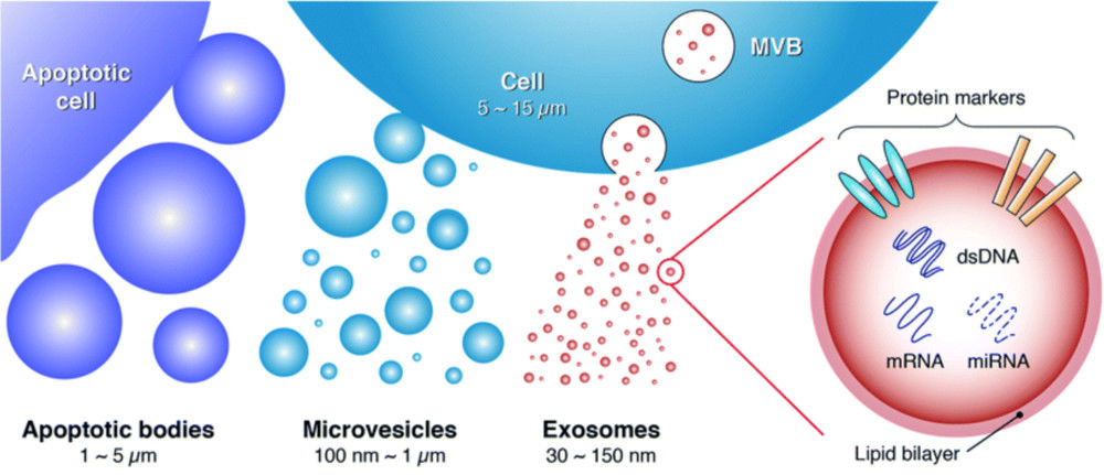
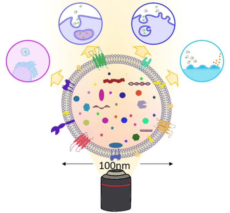
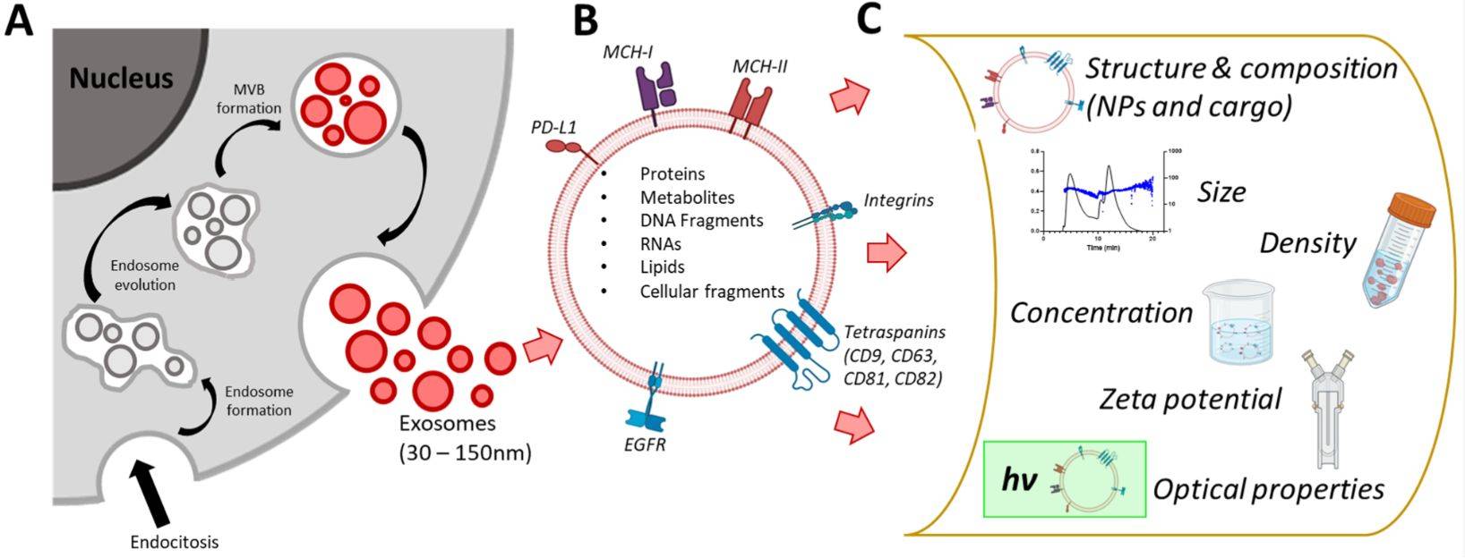
Exosomes are nanoscale extracellular vesicles (EVs) typically ranging from 30-150 nm in diameter. They carry bioactive molecules including proteins, lipids, and nucleic acids, acting as mediators of intercellular communication in cancer progression, immune regulation, and neurological disorders. With the growing clinical and industrial interest in exosome-based diagnostics and therapeutics, accurate and reproducible biophysical characterization has become essential.
Creative Biostructure offers a tunable resistive pulse sensing (TRPS) exosome characterization service that enables single-particle resolution and multiparametric analysis, delivering high-confidence data for both research and translational applications.
What is Tunable Resistive Pulse Sensing (TRPS)?
TRPS is an advanced implementation of Resistive Pulse Sensing (RPS), a technique based on the Coulter principle. In this method, particles suspended in an electrolyte pass through a nanopore under an applied electric field. Each particle transiently disrupts the ionic current, generating a "pulse" that is directly proportional to its size and influenced by its surface charge.
Unlike traditional RPS with fixed pores, TRPS employs stretchable, size-tunable nanopores, enabling precise adjustment of pore dimensions and voltage to analyze heterogeneous vesicle populations with higher accuracy. This flexibility makes TRPS especially suitable for exosome characterization, where size, concentration, and zeta potential are critical parameters.
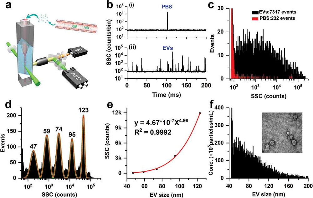
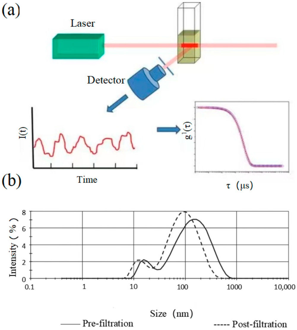
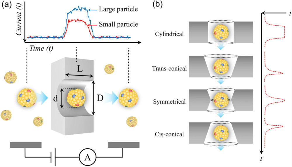 Figure 1. Schematic representation of resistive pulse sensing (RPS). (a) Measurement principle showing current changes as particles pass through a nanopore, generating resistive pulses. (b) Representative signal profiles reflecting pore morphology and particle translocation characteristics, enabling particle size and concentration analysis. (Kwon Y, et al., 2022)
Why Use TRPS for Exosome Analysis?
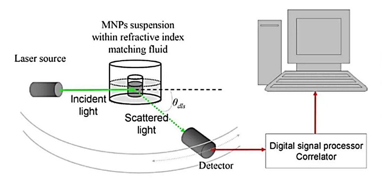
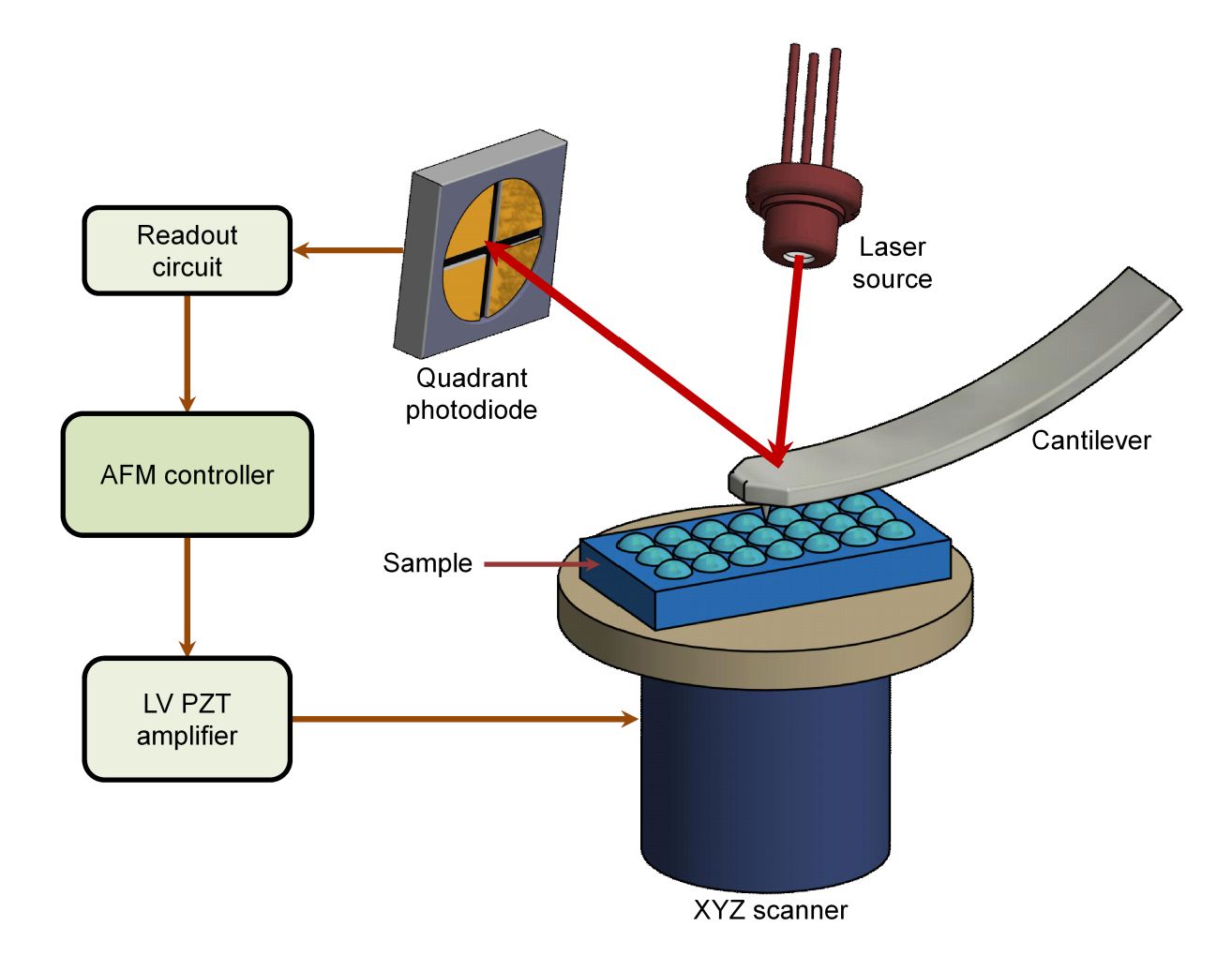
Conventional optical techniques such as Dynamic Light Scattering (DLS) and Nanoparticle Tracking Analysis (NTA) have well-documented limitations:
- DLS: Provides ensemble-averaged signals, often obscuring heterogeneity and unable to distinguish exosomes from protein aggregates.
- NTA: Offers particle tracking but may suffer from high variability (>30% error in concentration estimation) and lacks charge detection.
By contrast, TRPS offers true single-particle analysis, providing:
- Higher resolution of polydisperse samples.
- Absolute concentration measurement without reliance on standard curves.
- Capability to measure surface charge (zeta potential), which reflects membrane stability and intercellular interaction potential.
Comparison of DLS, NTA, and TRPS for Exosome Characterization
| Feature | DLS | NTA | TRPS |
|---|---|---|---|
| Principle | Measures light scattering from particle motion | Tracks scattered light of single particles | Detects current pulses as particles pass a nanopore |
| Resolution | Low (average, poor for mixed samples) | Medium (single particles, limited accuracy) | High (single-particle, precise size & charge) |
| Size Range | ~1 nm-6 μm | ~30-1000 nm | ~30-2000 nm |
| Concentration | Indirect, not reliable | Semi-quantitative, 20-50% error | Absolute count, <5% error |
| Zeta Potential | Bulk measurement only | Not available | Direct, single-particle |
| Sample Needs | High concentration, sensitive to aggregates | Moderate volume, works with fluids | Needs optimized buffer, risk of pore clogging |
| Strengths | Fast, simple, good for uniform samples | Size distribution & visualization | Most accurate for size, count, and charge |
| Limitations | Poor for heterogeneous samples | Lower reproducibility, errors in count | More complex, needs calibration & prep |
| Best Use | Quick screening | General exosome sizing | Detailed, quantitative exosome analysis |
Our TRPS Characterization Capabilities
Creative Biostructure provides comprehensive analysis of exosome samples using optimized TRPS workflows. Key measurable parameters include:
- Size Distribution: Nanoparticle diameters measured on a particle-by-particle basis, capturing heterogeneity across subpopulations.
- Concentration: Accurate quantification of vesicle numbers per unit volume.
- Surface Charge (Zeta Potential): Determination of electrophoretic mobility, reflecting vesicle stability, aggregation tendencies, and potential targeting properties.
We support a wide range of sample types, including:
- Cell culture supernatants
- Human and animal biofluids (serum, plasma, cerebrospinal fluid, urine)
- Purified exosome preparations from isolation workflows (ultracentrifugation, SEC, TFF, etc.)
Our system covers a dynamic size range from ~30 nm up to 2000 nm, enabling simultaneous detection of exosomes, microvesicles, and apoptotic bodies.
Our TRPS Exosome Characterization Workflow
At Creative Biostructure, we provide a comprehensive and standardized workflow for TRPS-based exosome characterization, ensuring every project is conducted with scientific rigor and compliance with MISEV2023 recommendations. From the initial consultation to the delivery of publication-ready reports, our process is designed to maximize data quality, reproducibility, and client confidence.
Project Consultation & Study Design
Project managers collaborate with clients to assess sample origin and research goals, then design tailored experimental plans with preparation guidance and defined deliverables.
Sample Receipt & Pre-Analytical Preparation
Clients may submit purified exosomes or request our exosome isolation service; we then verify sample quality, remove large particles, optimize dilution buffers, and document all pre-analytical steps for transparency.
TRPS Calibration & Instrument Setup
Calibration is performed with traceable standard beads, followed by adjustment of pore size, stretch, voltage, and pressure to match the target vesicle range, with all instrument details and calibration data documented in line with MISEV2023 guidelines.
Exosome Characterization
We provide real-time single-particle size profiling, absolute concentration counts without external standards, and zeta potential analysis to assess vesicle stability and interaction potential.
Quality Controls
Buffer-only runs define background signals, detergent-lysed controls confirm EV-specific events, and replicate measurements ensure reproducibility and robustness.
Data Analysis & Reporting
We deliver size, concentration, and zeta potential profiles with optional cross-validation, provide a detailed, publication-ready report, and offer expert consultation, downstream recommendations, and long-term project support.
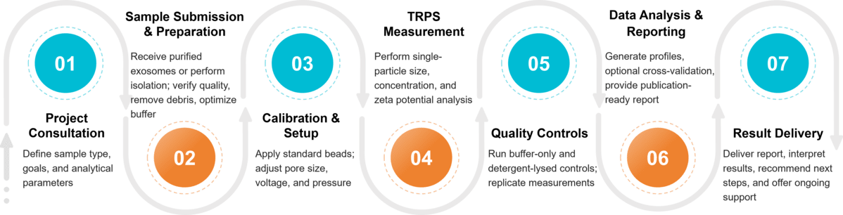 Figure 2. Project Workflow for Exosome Characterization by TRPS. (Creative Biostructure)
Sample Requirements for TRPS Measurement
To ensure accurate and reproducible TRPS analysis, Creative Biostructure recommends the following sample requirements. For rare or precious samples, customized solutions are available upon request.
| Requirement | Recommended Standard | Notes |
|---|---|---|
| Sample Volume | ≥100-200 μL | Minimum volume depends on concentration; low-volume protocols available for rare samples (≥50 μL). |
| Particle Concentration | ≥1 × 10⁹ particles/mL (optimal: 109-1010) | Lower concentrations may require enrichment or extended measurement time. |
| Sample Type | Purified exosomes or raw biofluids | Clients may submit pre-purified exosomes; isolation services available if needed. |
| Purity | Free of large debris, aggregates, or apoptotic bodies | Pre-clarification (filtration/centrifugation) recommended to prevent pore clogging. |
| Buffer Conditions | PBS or other low-conductivity buffers | Surfactant additives may be used to minimize aggregation and stabilize pore performance. |
| Replicates | ≥3 runs per sample | Ensures reproducibility and statistical robustness of results. |
What Deliverables Will You Receive
- Size distribution histograms with single-particle resolution
- Absolute concentration statistics (particles/mL)
- Zeta potential profiles indicating vesicle stability
- Full technical report with workflow, QC details, and instrument settings
Applications of TRPS in Exosome Research
- Exosome-based drug development: Monitor particle size uniformity and surface charge of engineered or drug-loaded vesicles to ensure product consistency.
- Biomarker discovery: Identify pathological changes in exosome subpopulations associated with cancer, neurodegeneration, or immune dysfunction.
- Fundamental EV biology: Explore correlations between size/charge heterogeneity and biological function.
- Quality control for exosome manufacturing: Verify batch reproducibility in preclinical and GMP workflows.
Why Choose Creative Biostructure?
- Trusted Expertise: 10+ years in exosome and structural biology analysis.
- High Accuracy: Optimized TRPS workflows with cross-validation by TEM, NTA, and more.
- Comprehensive Solutions: From isolation to multiparametric characterization and reporting.
- Client-Centered Support: Dedicated consultation and tailored recommendations for every project.
Case Study
Case: TRPS-Based Characterization of USC-Derived Exosomes in a Rett Syndrome Model
Background
Rett syndrome (RTT) is a neurodevelopmental disorder linked to MECP2 mutations. Human urine-derived stem cell exosomes (USC-Exos), enriched in miR-21-5p, were studied for their neurogenic potential via the EphA4/TEK pathway. Precise vesicle characterization was critical.
Methods
Exosomes were analyzed by TEM, western blot, and TRPS using the qNano platform with NP100 nanopores and CPC100 calibration beads.
Results
TRPS showed USC-Exos were mainly 50-100 nm with a concentration of 5.1 × 10¹⁰ particles/mL, consistent with TEM findings. Marker profiling confirmed purity. Functionally, miR-21-5p-rich exosomes enhanced neuronal differentiation and improved motor and cognitive function in RTT mice.
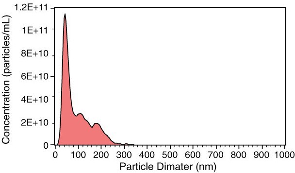 Figure 3. TRPS Measurement of USC-Exo Size and Concentration. (Pan W, et al., 2021)
Conclusion
TRPS provided high-resolution size and concentration data essential for validating exosome identity and purity, ensuring reliable biological interpretation.
At Creative Biostructure, we are committed to delivering accurate, reproducible, and publication-ready exosome data through our advanced TRPS platform. Whether your project focuses on biomarker discovery, therapeutic exosome development, or quality control, our expert team is ready to support you with tailored solutions. Contact us to discuss your project and request a customized quote.
References
- Pan W, Xu X, Zhang M, et al. Human urine-derived stem cell-derived exosomal miR-21-5p promotes neurogenesis to attenuate Rett syndrome via the EPha4/TEK axis. Laboratory Investigation. 2021, 101(7): 824-836.
- Kwon Y, Park J. Methods to analyze extracellular vesicles at single particle level. Micro and Nano Systems Letters. 2022, 10(1): 14.
- Welsh J A, Goberdhan D C I, O'Driscoll L, et al. Minimal information for studies of extracellular vesicles (MISEV2023): From basic to advanced approaches. Journal of Extracellular Vesicles. 2024, 13(2): e12404.
Frequently Asked Questions
For any inquiries, our support team is ready to help you get technical support for your research and maximize your experience with Creative Biostructure.