Exosome Bioluminescent Labeling Services
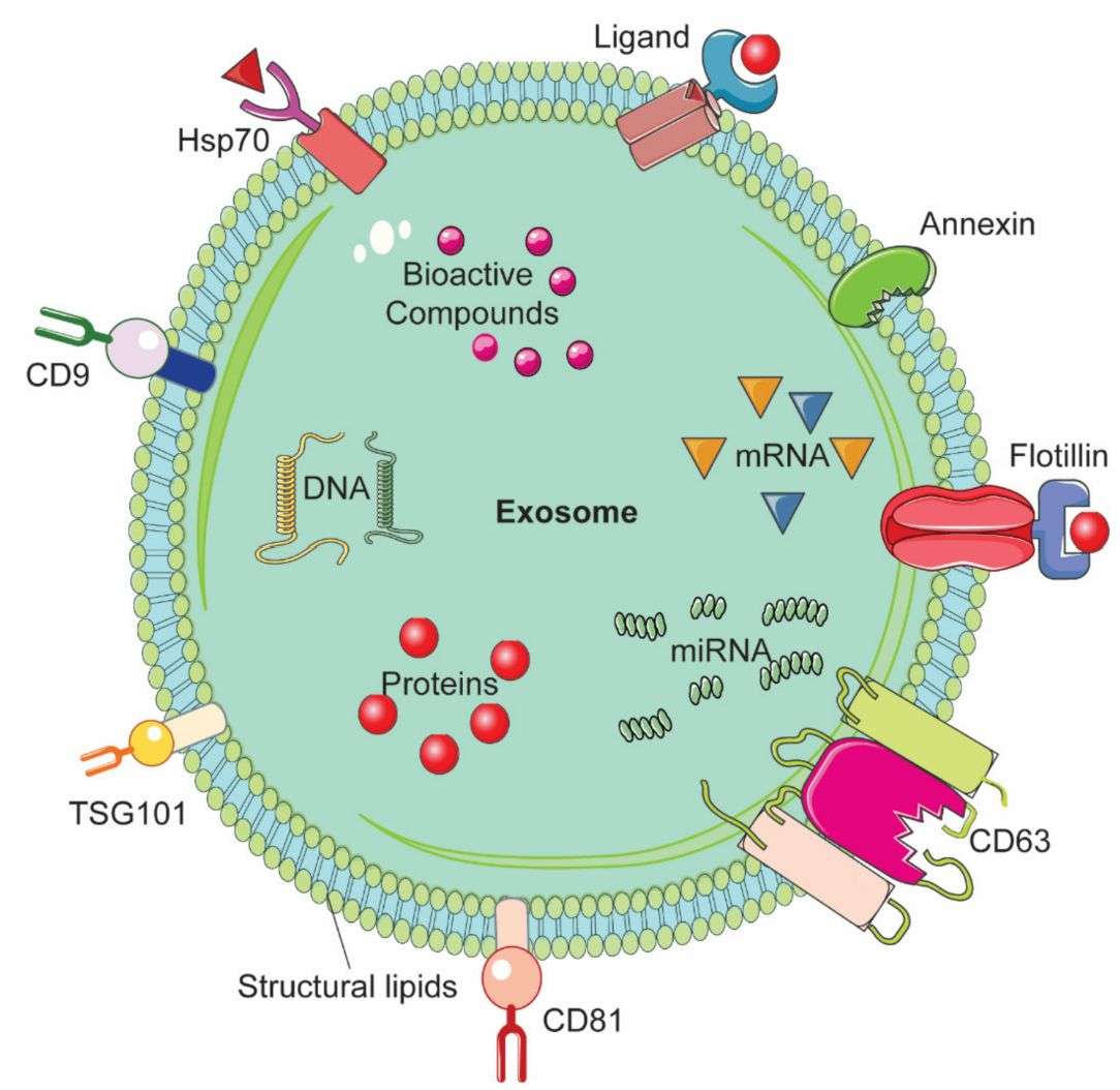
Bioluminescent labeling enables ultrasensitive, non-invasive tracking of exosomes in vitro and in vivo through luciferase reporters fused to exosomal markers such as CD63, CD9, or CD81, generating quantifiable light signals upon substrate addition without external excitation. Creative Biostructure provides end-to-end exosome bioluminescent labeling services, including construct design, cell engineering, labeled exosome production, and characterization for reliable biodistribution and functional studies.
Why Bioluminescent Labeling Matters in Exosome Research
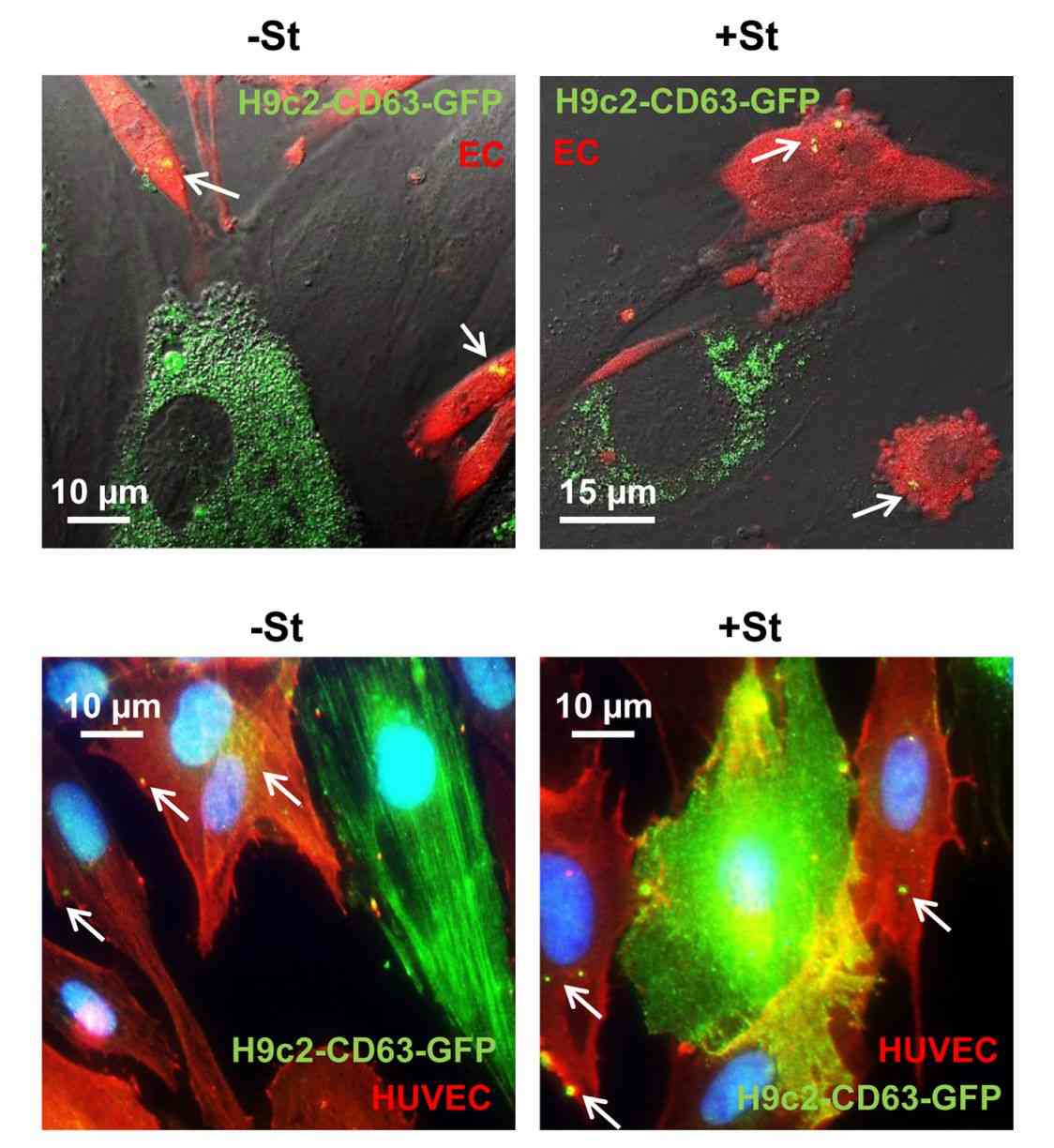
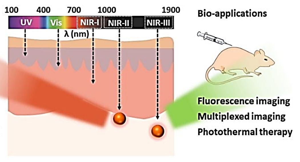
Fluorescence-based tracking is constrained by autofluorescence, photobleaching, and limited tissue penetration from external excitation. Bioluminescence imaging overcomes these limits by producing light via luciferase–substrate reactions, enabling zero-background, highly sensitive, and more directly quantitative exosome detection.
Advances such as NanoLuc and BRET-enabled systems further improve sensitivity and extend imaging into deeper tissues.
Key advantages:
- Zero-background signal without excitation light
- Direct quantitative correlation with exosome abundance
- High sensitivity for low-abundance and near-single-vesicle detection
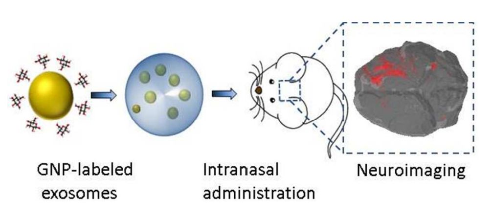
- Non-invasive longitudinal in vivo tracking
- ATP-independent luminescence for stable extracellular signals
- BRET-based near-infrared emission for deep-tissue imaging
- Multiplexed tracking of distinct exosome populations.
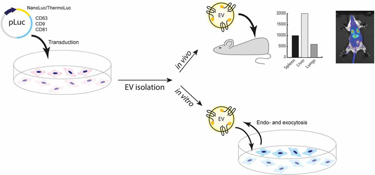 Figure 1. Bioluminescent Imaging of Extracellular Vesicle Biodistribution. (Gupta D, et al., 2020)
Bioluminescent vs. Fluorescent Labeling
While both modalities enable exosome tracking, their suitability depends on the research question and experimental design.
| Parameter | Bioluminescent Labeling | Fluorescent Labeling | Preferred Application Scenario | Key Limitation |
|---|---|---|---|---|
| Light source | Enzymatic (no excitation) | External laser/LED | Deep tissue, longitudinal in vivo BLI | Requires genetic engineering of producer cells |
| Background signal | Essentially zero | Variable (autofluorescence) | Quantitative detection of low-abundance exosomes | Substrate cost for repeated imaging |
| Signal type | Quantitative (particle-proportional) | Semi-quantitative (variable per dye) | Exosome secretion rate measurement | Dependence on substrate pharmacokinetics |
| Multiplexing | Substrate-selective luciferase pairs | Spectrally distinct fluorophores | Simultaneous dual-population tracking | Limited spectral channels vs. fluorescence |
| In vivo depth | BRET-NIR: >1 cm; NLuc: ~5 mm | NIR: ~1 cm; Visible: ~1-2 mm | Whole-body BLI and deep-organ imaging | NLuc alone (~460 nm) absorbed by hemoglobin |
| Sample processing | Direct luminescence readout of medium | Requires dye removal and washing | High-throughput screening | Requires stable producer cell line |
Our Bioluminescent Exosome Labeling Service Portfolio
Creative Biostructure provides end-to-end luciferase-based exosome labeling solutions. Each project starts with feasibility assessment to align reporter selection, construct design, cell engineering, and imaging strategy with downstream application needs.
1. CD63/CD9/CD81–NanoLuc Exosome Production (ExoLuc System)
We engineer producer cell lines to stably express NanoLuc fused to exosome-enriched markers (CD63, CD9, or CD81), generating highly sensitive bioluminescent exosomes for quantitative and in vivo studies.
Service scope:
- Lentiviral or genome-integrated expression of NLuc-tetraspanin constructs
- Stable clone screening and expression validation
- Exosome isolation (UC + SEC) and physicochemical characterization
- Luminescence-based QC and batch consistency testing
- Delivery of purified, fully characterized NLuc-labeled exosomes
2. Gaussia Luciferase Dual-Reporter Exosome System
We generate engineered exosome-producing cells expressing targeted gLuc fused with fluorescent reporters, enabling dual-modality imaging and orthogonal validation of exosome release and uptake.
Service scope:
- Design of exosome-targeted gLuc-fluorescent fusion constructs
- Stable cell line generation and secretion validation
- Functional pathway confirmation using secretion inhibition controls
- Dual readouts: bioluminescence, fluorescence imaging, and EV characterization (NTA/TEM)
3. BRET-Enhanced Near-Infrared Exosome Labeling
We develop BRET-based luciferase systems that shift emission into red or near-infrared ranges for improved in vivo imaging depth and signal resolution.
Service scope:
- NLuc donor paired with red/NIR acceptors or dye conjugation systems
- Linker engineering to optimize energy transfer efficiency
- Spectral profiling and BRET ratio optimization
- In vivo imaging validation with whole-animal IVIS analysis
4. Custom Luciferase-Fusion Exosome Engineering
We offer fully customized reporter design for specialized applications, including multiplex EV tracking, targeted delivery studies, and tissue-specific exosome labeling systems.
Service scope:
- Custom construct design and codon optimization
- Transient or stable expression system development
- Functional validation (incorporation, kinetics, signal stability)
- Scalable production from pilot to preclinical-grade batches
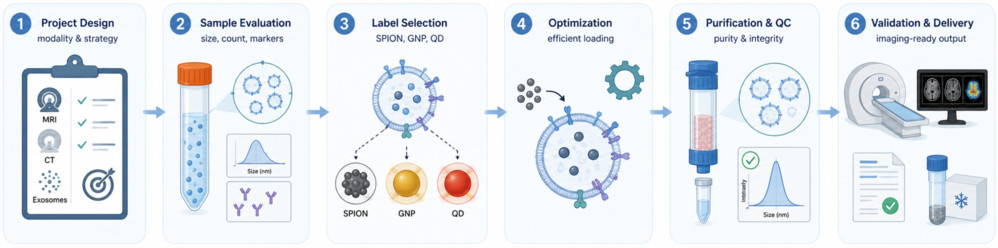
Standard Workflow for Bioluminescent Exosome Labeling
- Study Design: Define exosome source, cell system, and imaging mode (in vitro BLI or in vivo IVIS); select luciferase strategy.
- Construct Design: Build luciferase fusions (NLuc, gLuc, BRET, or custom) with optimized promoters and linkers.
- Cell Engineering: Generate stable producer cells via lentiviral transduction or genome editing, followed by clonal selection.
- Clone Validation: Screen for luciferase activity and confirm correct exosomal incorporation.
- Exosome Production: Expand validated clones and isolate exosomes via UC + SEC or TFF workflows.
- QC & Delivery: Perform luminescence calibration, NTA/TEM characterization, marker validation, and deliver QC-backed exosome batches.
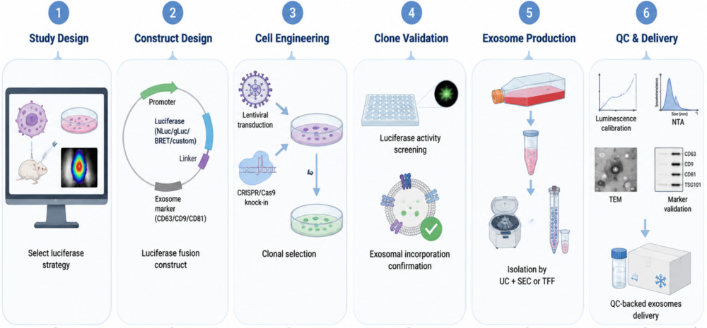 Figure 1. Standard Workflow for Bioluminescent Exosome Labeling. (Creative Biostructure)
Characterization and Quality Control Parameters
Each batch of bioluminescent-labeled exosomes is subjected to a standardized multi-parameter QC workflow to ensure consistency, purity, and functional signal reliability.
- Size & concentration: NTA (±DLS as needed)
- Morphology: TEM (cryo-EM available on request)
- Luminescence performance: Serial dilution assay with RLU-particle correlation
- Signal kinetics: Time-course profiling to define optimal imaging window
- Protein validation: Western blot for luciferase fusion and exosome markers (CD9, CD81, TSG101, Alix)
- Signal specificity: Comparison with unlabeled controls at matched particle counts
- Purity check: SEC-based confirmation of luciferase co-elution with exosome fractions
- Stability (optional): Luminescence retention at 4°C, −20°C, −80°C over time
- Safety testing (in vivo grade): Endotoxin (LAL) and sterility assessment
Project Initiation Options
| Option | Description | What You Provide | What We Deliver |
|---|---|---|---|
| Customer-Provided Cell Line | We transduce your cell line with the luciferase-fusion construct and produce labeled exosomes. | Viable cell line + project brief | Stable transduced cell line + labeled exosomes + QC report |
| Full-Service (End-to-End) | We handle construct design, cell engineering, exosome production, and characterization. | Project requirements and target specifications | Full production records + labeled exosomes + comprehensive QC report |
| Construct-Only Service | We design and clone the luciferase-fusion construct; you perform cell engineering and exosome production. | Target gene sequence or construct specifications | Validated plasmid construct + sequence verification + cloning report |
What Deliverables Will You Receive
| Deliverable | Details |
|---|---|
| Exosome Product | Purified luciferase-labeled exosomes in defined buffer at specified concentration |
| Exosome Characterization | Size distribution (D10/D50/D90), particle concentration, and TEM images confirming morphology |
| Functional Assays | Luminescence profiling (RLU titration, kinetics, signal linearity) |
| Molecular QC | Western blot for luciferase fusion and exosome markers (CD9, CD81, TSG101); SEC-based free luciferase removal |
| Construct QC (if applicable) | Sequence verification of engineered constructs |
| Documentation | Integrated technical report including protocols, raw data, and QC interpretation |
| Support | Optional downstream imaging and data analysis support |
Why Choose Creative Biostructure for Bioluminescent Exosome Labeling
- Broad luciferase expertise: NLuc, gLuc, FLuc, and BRET systems tailored to different imaging needs
- End-to-end solution: From construct design and cell engineering to purified labeled exosome delivery
- Application-matched design: System selection optimized for imaging depth, sensitivity, and quantitative requirements
- Reliable data quality: Built-in controls ensure signal specificity, linearity, and removal of free enzyme background
- Clear, publication-ready output: Complete QC documentation with full data package on request
Case Study
Case: In Vivo Tracking of Tumor-Derived Exosomes Using CD63-NanoLuc Labeling
Background
Tracking endogenous exosomes in vivo is challenging due to their nanoscale size and rapid systemic distribution. This study applied a CD63-NanoLuc bioluminescent system to enable real-time monitoring of tumor-derived exosomes during early DCIS progression.
Methods
- Labeling strategy: CD63-NanoLuc fusion for exosome-specific bioluminescence
- Model: Orthotopic DCIS mouse intraductal xenograft model
- Readouts: In vivo IVIS imaging, ex vivo organ imaging, plasma assays, molecular validation (qPCR, RNA-seq, IHC)
Results
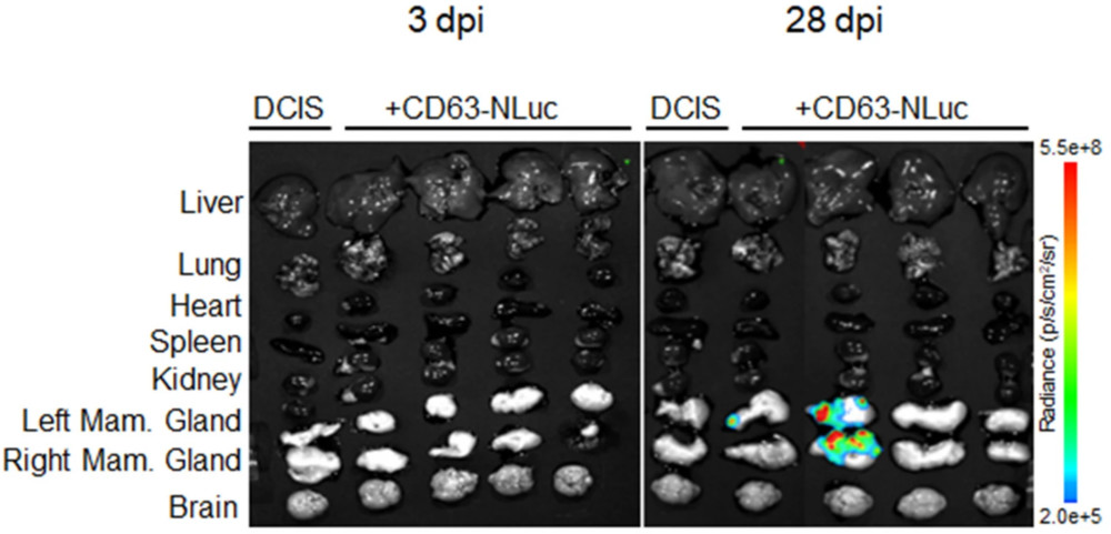 Figure 3. Ex vivo bioluminescence imaging reveals time-dependent, organ-specific distribution of CD63-NanoLuc-labeled exosomes in a DCIS mouse model, with signal intensities increasing across multiple tissues compared with controls. (Hladik C, et al., 2026)
- Early detection: Exosome signals detected by ~day 14 post-implantation and increased over time
- Systemic spread: Signals observed in multiple organs including liver, lung, spleen, brain, and heart
- Circulation evidence: Plasma NLuc activity detected prior to overt invasion
- Organ specificity: Kidney showed minimal signal, indicating selective biodistribution
- Origin confirmation: Absence of NLuc mRNA in distant tissues confirmed exosome-derived signal
- Systemic effects: Brain transcriptome changes suggested early distant microenvironment modulation
Conclusion
CD63-NanoLuc labeling enables sensitive, longitudinal in vivo tracking of tumor-derived exosomes, revealing early systemic dissemination and organ-specific biodistribution during DCIS progression.
Ready to track exosome secretion, biodistribution, and inter-organ communication with zero-background, quantitative bioluminescent sensitivity? Contact us to discuss the optimal luciferase system, construct design, and cell engineering strategy for your research. Our team provides end-to-end support from molecular cloning to publication-ready labeled exosome delivery.
References
- Gupta D, Liang X, Pavlova S, et al. Quantification of extracellular vesicles in vitro and in vivo using sensitive bioluminescence imaging. Journal of Extracellular Vesicles. 2020, 9(1): 1800222.
- Hladik C, Elayapillai S P, Dogra S, et al. Nanoluciferase-CD63 labeling reveals extracellular vesicle kinetics in a mouse intraductal model of ductal carcinoma in situ. Breast Cancer Research. 2026.
Frequently Asked Questions
For any inquiries, our support team is ready to help you get technical support for your research and maximize your experience with Creative Biostructure.