Exosome Radiolabeling Services
Exosome radiolabeling enables highly sensitive SPECT/PET imaging for quantitative in vivo tracking of biodistribution, pharmacokinetics, and therapeutic behavior. Compared with optical methods, it provides deep-tissue penetration and absolute signal quantification. Creative Biostructure delivers end-to-end exosome radiolabeling services, including isotope selection, labeling optimization, purification, quality control, and imaging support. We apply tailored strategies such as chelator-based labeling, intraluminal labeling, and radioiodination to ensure stable, high-specific-activity, and reproducible imaging results.
What Is Exosome Radiolabeling for SPECT/PET Imaging
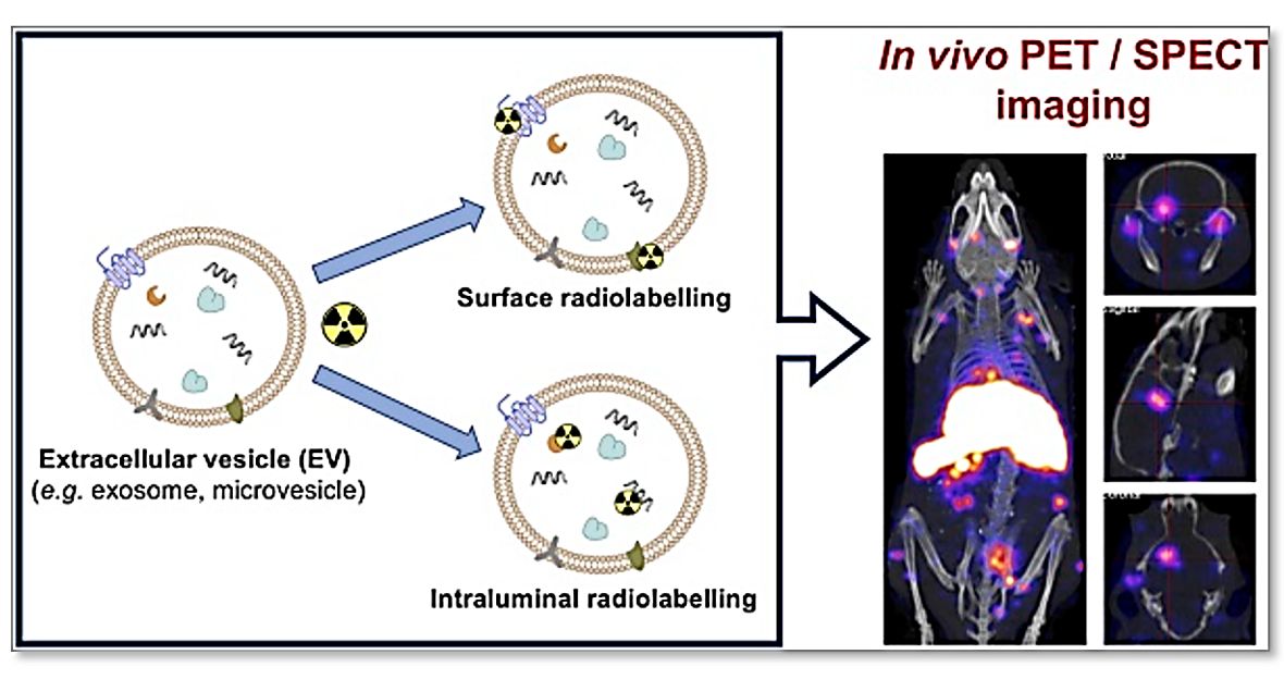
Exosome radiolabeling for SPECT/PET imaging refers to the incorporation of gamma- or positron-emitting radionuclides into or onto exosomes, enabling non-invasive detection of their distribution in living animals. Two principal radiolabeling strategies are used in contemporary EV research:
- Indirect radiolabeling: A bifunctional chelator (e.g., DFO, NOTA, DOTA) is conjugated to exosome surface proteins or lipids, followed by coordination with metal radionuclides (e.g., 89Zr, 64Cu, 68Ga).
- Direct radiolabeling: Lipophilic radiotracers (e.g., [89Zr]Zr(oxinate)4, 99mTc-HMPAO) passively diffuse across the exosome membrane and are retained in the lumen or membrane, without requiring chemical conjugation.
Why Radiolabeling Matters for In Vivo Exosome Tracking
Optical imaging modalities (fluorescence, bioluminescence) are constrained by tissue autofluorescence, photon scattering, and limited penetration depth (<1 mm), making them unsuitable for deep-tissue or whole-body biodistribution studies in vivo. Nuclear imaging overcomes these limitations:
- Deep-tissue penetration: Gamma photons (SPECT) and 511-keV annihilation photons (PET) enable non-invasive whole-body imaging.
- Absolute quantification: Regions of interest (ROIs) can be converted to %ID/g (percentage of injected dose per gram tissue) using calibration standards.
- High sensitivity: Picomolar-to-femtomolar detection enables tracking of low-dose exosome administrations relevant to translational studies.
- Temporal resolution: Longitudinal imaging in the same animal over hours to days reduces inter-animal variability and overall animal use.
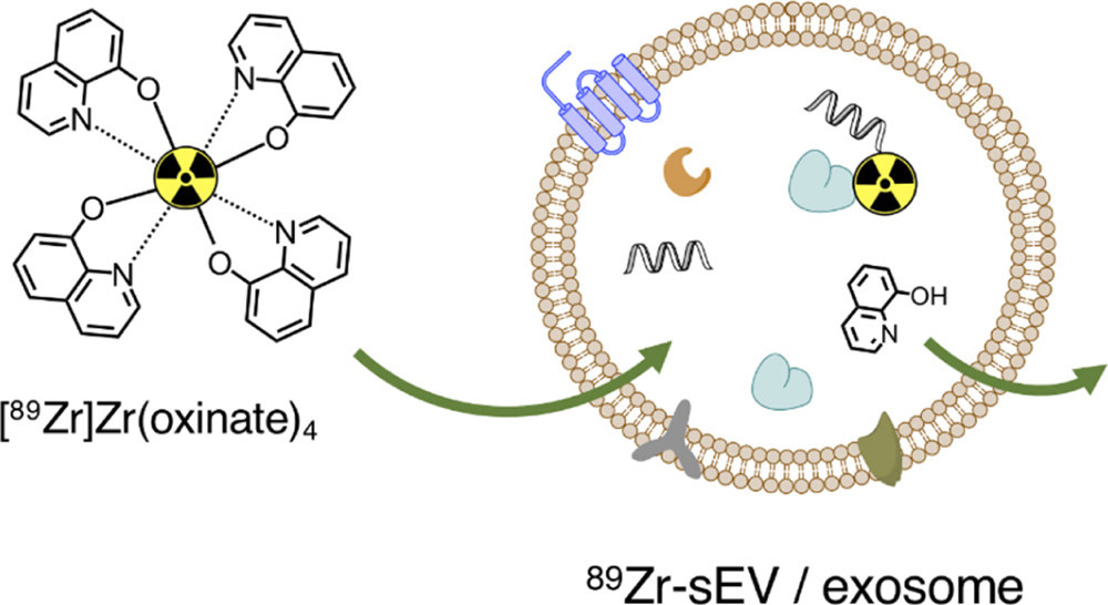 Figure 1. Intraluminal 89Zr Radiolabeling Mechanism of Small Extracellular Vesicles (sEVs). (Khan A A, et al., 2022)
Our Advanced Exosome Radiolabeling Services for SPECT/PET
Creative Biostructure provides a full suite of radiolabeling services tailored to the isotope, exosome source, and imaging objective. Each project is scoped through a pre-labeling consultation to match the radionuclide and labeling chemistry to the preclinical question, the imaging modality (SPECT or PET), and the required temporal window.
Radionuclide and Labeling-Strategy Portfolio
| Radionuclide | Imaging Modality | Half-Life | Key Labeling Method | Primary Applications | Special Considerations |
|---|---|---|---|---|---|
| 99mTc | SPECT | 6.0 h | HMPAO, direct reduction | Acute biodistribution, organ uptake (liver/spleen) | Same-day imaging; low cost; widespread availability |
| 111In | SPECT | 2.8 d | Oxine, DTPA | Multi-day SPECT tracking, ex vivo biodistribution | Long half-life; potential for 111In-transferrin transchelation |
| 124I | PET | 4.2 d | Bolton-Hunter, IODO-GEN | Multi-day PET; thyroid blocking required | Block thyroid with KI; 124I also emits gamma |
| 89Zr | PET | 78.4 h | DFO-chelation, [89Zr]Zr(oxinate)4 | Multi-day PET, stem cell-derived exosomes | DFO conjugation mild; oxinate method is direct and fast |
| 64Cu | PET | 12.7 h | NOTA, DOTA | Same-day to next-day PET; theranostic pairing | 64Cu has β+ fraction ~17.6%; NOTA preferred for stability |
| 18F | PET | 109.8 min | ALT, SFB, click chemistry | High-resolution small-animal PET | Requires rapid conjugation; ultra-high sensitivity |
Labeling Strategies We Offer
- Indirect chelation labeling (DFO-89Zr, NOTA-64Cu, DOTA-68Ga): Surface conjugation of bifunctional chelators to exosome proteins or amino lipids, followed by radiometal coordination. Optimized conditions preserve vesicle integrity and achieve >90% radiochemical purity.
- Direct intraluminal radiolabeling ([89Zr]Zr(oxinate)4): Passive diffusion of the lipophilic 89Zr-oxinate complex into the exosome lumen, requiring no chemical conjugation and completing within 15-30 min at room temperature.
- 99mTc-HMPAO radiolabeling: Reduction of 99mTc-pertechnetate with stannous chloride in the presence of HMPAO, followed by incubation with exosomes. The lipophilic 99mTc-HMPAO complex crosses the membrane and is retained.
- Radioiodination (124I/125I): Indirect labeling of surface tyrosine residues using Bolton-Hunter reagent or IODO-GEN oxidation. Suitable for PET (124I) and SPECT/ex vivo (125I) imaging.
- Dual-isotope labeling: Concurrent or sequential incorporation of two isotopes (e.g., 99mTc for SPECT and 89Zr for PET) for cross-validation or multi-parameter tracking.
- Custom radiochemistry: For novel isotopes, chelators, or click-chemistry approaches, we perform feasibility assessment and protocol optimization using the client's exosome system.
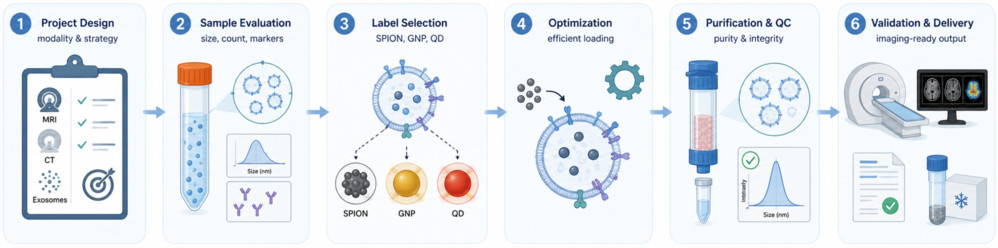
Our End-to-End Radiolabeling Workflow
- Consultation & isotope selection: Define study goals and select optimal radionuclide and labeling strategy based on exosome source and imaging needs.
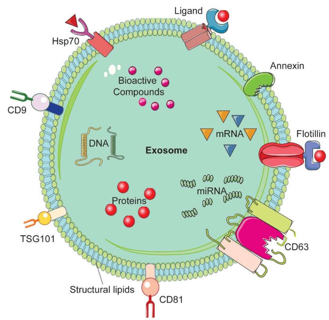
- Exosome characterization: Pre-labeling assessment of size/concentration (NTA), morphology (TEM), and markers (CD9/CD63/CD81).
- Radiolabeling reaction: Isotope-specific labeling under ALARA-compliant, shielded conditions to preserve exosome integrity.
- Purification & RCP analysis: Removal of free isotope via SEC/filtration/dialysis, with radiochemical yield and purity confirmation (TLC/SEC).
- QC & stability testing: Evaluation of particle integrity and in vitro stability; optional sterility and endotoxin testing for in vivo use.
- In vivo support & reporting: Guidance on dosing, imaging, ROI analysis, %ID/g quantification, and structured data reporting.
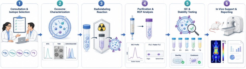 Figure 1. Exosome Radiolabeling Workflow for SPECT/PET Imaging. (Creative Biostructure)
Comprehensive Quality Control and Characterization
- Labeling efficiency: Evaluated by TLC or SEC with fraction analysis and radioactivity measurement.
- Radiochemical purity (RCP): Typically >90% at release, with follow-up assessment in serum to verify short-term stability.
- Particle size & concentration: NTA pre- and post-labeling to monitor size distribution changes and aggregation risk.
- Morphology: TEM/cryo-EM used to confirm structural integrity of vesicles after labeling.
- Specific activity: Reported as activity per particle number or protein amount to support dosing.
- In vitro stability: Serum incubation at physiological temperature with time-course monitoring of label retention.
- Sterility & endotoxin: Standard testing applied for preparations intended for in vivo use.
Applications (Research Use Only)
- Whole-body biodistribution studies of systemically administered exosomes in mice, rats, or larger animal models.
- Organ tropism mapping (liver, spleen, lung, kidney, brain, tumor) to support dose and route selection.
- Pharmacokinetic profiling, including blood half-life, AUC, and clearance pathways (renal, hepatobiliary).
- Comparative biodistribution of engineered vs. native exosomes (e.g., modified, loaded, or ligand-decorated).
- Longitudinal tracking of the same animal over days to weeks.
- Specificity evaluation using blocking or competitive uptake studies.
- Translational pharmacokinetic studies supporting IND-enabling preclinical packages.
What Deliverables Will You Receive
| Deliverable | Details |
|---|---|
| Radiolabeled product | Sterile, imaging-ready radiolabeled exosomes (defined activity/volume) |
| Radiolabeling report | Isotope, labeling efficiency, RCP (0 h & stability check), specific activity |
| QC characterization | NTA (size/concentration), optional TEM and marker validation |
| Purity verification | TLC/SEC-based radiochemical purity data |
| Imaging data (if applicable) | Raw DICOM/NIfTI files and %ID/g ROI quantification |
| Technical documentation | Full methods, results summary, and handling/storage guidance |
| Optional analysis | Pharmacokinetic modeling (AUC, half-life, compartment analysis) |
How to Get Started
Clients may provide purified exosomes or request upstream isolation and production support. Project initiation follows a streamlined workflow:
- Share your exosome source, target radionuclide (or request recommendation), imaging modality, and animal model.
- Our team assesses feasibility, advises on isotope logistics, and provides a tailored proposal with timeline and cost.
- Upon approval, we perform radiolabeling and QC under controlled conditions and deliver the radiolabeled product with full documentation and imaging support as needed.
Why Choose Creative Biostructure
- Radiochemistry expertise: Extensive experience with 99mTc, 89Zr, 64Cu, 124I, and 18F labeling of extracellular vesicles.
- Rigorous QC standards: Multi-parameter release criteria (RCP >90%, size stability, morphology) ensure reproducible data quality.
- Isotope-flexible design: Support for SPECT and PET tracers with guidance on optimal isotope selection.
- Translational focus: Workflows aligned with GLP/GMP principles to support progression toward clinical studies.
- Integrated services: Radiolabeling combined with exosome isolation, characterization, and engineering in one workflow.
- Regulatory compliance: Full adherence to radiation safety standards, with support for institutional approvals when required.
Case Study
Case: Tc-99m Radiolabeled Exosome SPECT Imaging (UCMSC-EVs)
Background
UCMSC-derived exosomes were radiolabeled with Tc-99m to enable quantitative SPECT imaging for in vivo biodistribution and pharmacokinetic analysis.
Methods
- Labeling: Direct SnCl2-mediated Tc-99m surface radiolabeling
- Model: BALB/c mice (IV injection)
- Imaging: SPECT/CT + ex vivo gamma counting
- Analysis: iTLC, HPLC, and two-compartment PK modeling
Results
- Radiolabeling: ~52% yield, >99% purity
- Stability: Maintained up to 24 h in serum
- Integrity: No significant change in size or exosomal markers
- Biodistribution: Predominant uptake in liver and spleen
- PK: Rapid distribution (0.85 min) and elimination (~25 min)
- Clearance: Primarily urinary excretion
Conclusion
Tc-99m labeling enables stable, quantitative tracking of exosomes in vivo, supporting reliable biodistribution and pharmacokinetic assessment for translational research.
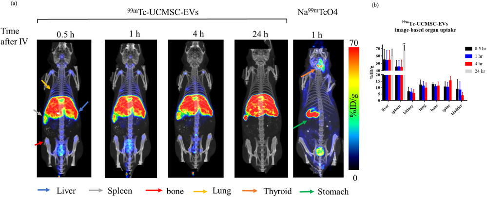 Figure 3. Representative SPECT/CT imaging and quantitative biodistribution of Tc-99m-labeled UCMSC-derived exosomes compared with free pertechnetate control over time. Radiolabeled exosomes show predominant accumulation in liver and spleen, minimal thyroid/stomach signal, and urinary clearance. (Chung Y H, et al., 2024)
Ready to quantify your exosome biodistribution with SPECT or PET imaging? Contact us to discuss isotope selection, radiolabeling strategy, imaging coordination, and a customized project plan.
References
- Khan A A, Man F, Faruqu F N, et al. PET imaging of small extracellular vesicles via [89Zr] Zr (oxinate) 4 direct radiolabeling. Bioconjugate Chemistry. 2022, 33(3): 473-485.
- Chung Y H, Ho Y P, Farn S S, et al. In vivo SPECT imaging of Tc-99m radiolabeled exosomes from human umbilical-cord derived mesenchymal stem cells in small animals. Biomedical Journal. 2024, 47(5): 100721.
- Li J, Li C, Zheng H, et al. Biodistribution studies of 89Zr-labeled stem cell-derived exosomes using PET imaging. Applied Radiation and Isotopes. 2025, 225: 112000.
Frequently Asked Questions
For any inquiries, our support team is ready to help you get technical support for your research and maximize your experience with Creative Biostructure.