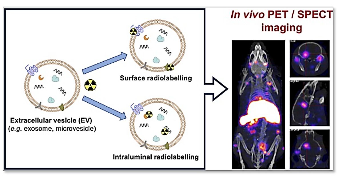
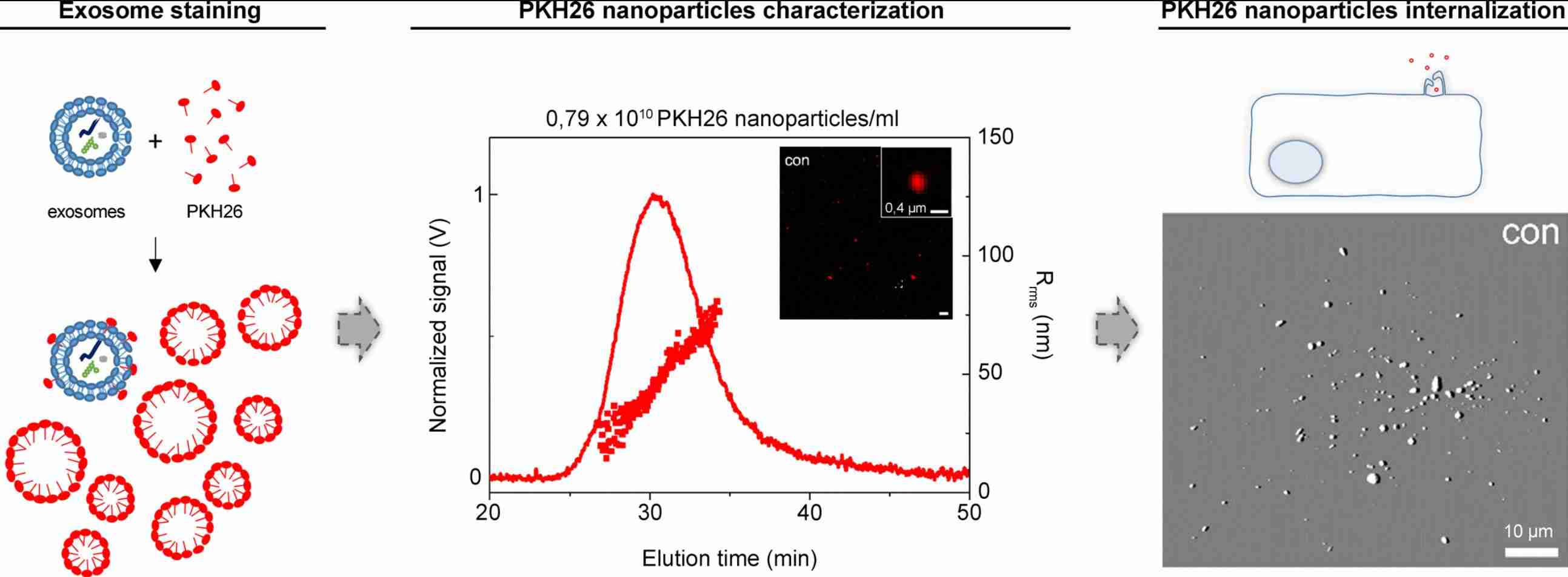
Exosome Magnetic and Contrast Agent Labeling Services
Creative Biostructure provides exosome magnetic and contrast agent labeling services for imaging, tracking, and quantification of exosomes beyond conventional fluorescence. Our portfolio includes SPION/USPIO labeling for MRI tracking, gold nanoparticle labeling for CT and electron-dense imaging, and quantum dot labeling for fluorescence and multiplexed uptake analysis. Each project is tailored to the exosome source and imaging platform, with optimized labeling, purification, and quality control workflows to minimize free-particle artifacts and preserve exosome integrity for reliable preclinical biodistribution and uptake studies.
Why Magnetic and Contrast Agent Labeling Matters in Exosome Research
Optical imaging modalities such as fluorescence and bioluminescence are widely used for exosome tracking but are limited by tissue scattering and shallow in vivo penetration. Magnetic and contrast imaging overcomes these limitations by enabling deep-tissue, whole-body detection through MRI and CT, supporting accurate exosome biodistribution studies in preclinical and translational models.
Key advantages of magnetic and contrast agent labeling:
- Deep tissue imaging: MRI and CT enable whole-body exosome tracking independent of penetration depth.
- Clinical relevance: SPIONs and gadolinium-based agents are established MRI contrast agents with proven translational use.
- Quantitative readout: MRI relaxation changes and CT attenuation correlate with contrast agent concentration for semi-quantitative analysis.
- High-resolution localization: MRI provides precise organ-level mapping of exosome distribution.
- Multi-modal compatibility: Magnetic labeling can be combined with optical or nuclear imaging for complementary validation.
Magnetic/Contrast Agent vs. Optical Labeling
While both optical and magnetic approaches are widely used in exosome research, the optimal choice depends on imaging depth, quantification needs, and translational relevance.
| Parameter | Magnetic/Contrast Agent Labeling | Optical Labeling (Fluorescence/BLI) | Preferred Application Scenario |
|---|---|---|---|
| Signal mechanism | Magnetic field or X-ray attenuation–based contrast | Photon emission (fluorescence or luciferase) | Deep-tissue vs. cellular-scale tracking |
| Tissue penetration | Unlimited (MRI/CT) | Limited (mm–cm range) | Whole-body biodistribution studies |
| Spatial resolution | MRI/CT: organ-level (μm–sub-mm) | Optical: cellular to mm-scale in vivo | Anatomical localization vs. cell-level imaging |
| Quantification | Signal correlates with contrast agent concentration (R2/R2*) | Semi-quantitative or relative intensity | Organ-level exosome burden estimation |
| Clinical translatability | High (clinically used contrast agents) | Low (preclinical use) | Translational and IND-enabling studies |
| Multiplexing | MRI-specific contrast channels (T1/T2/19F) | Multi-color fluorophores or luciferase systems | Multi-population tracking strategies |
Magnetic and Contrast Agent Labeling Strategies for Exosomes
Creative Biostructure provides exosome labeling services based on three core nanomaterial platforms. Each platform supports distinct imaging needs and experimental goals. Custom or multimodal labeling strategies can also be developed upon request.
SPION / USPIO Labeling
- Principle: Iron oxide nanoparticles enable MRI contrast via T2/T2* signal reduction
- Applications: Whole-body exosome biodistribution; organ-level tracking; clearance and homing studies
- Advantages: Deep-tissue MRI imaging; quantitative iron-based readout; clinically relevant imaging platform
- Key validation: Loading efficiency; free vs. encapsulated iron quantification; aggregation and exosome integrity assessment; MRI phantom pre-validation
Gold Nanoparticle (GNP) Labeling
- Principle: Gold nanoparticles generate CT contrast and electron-dense TEM signals
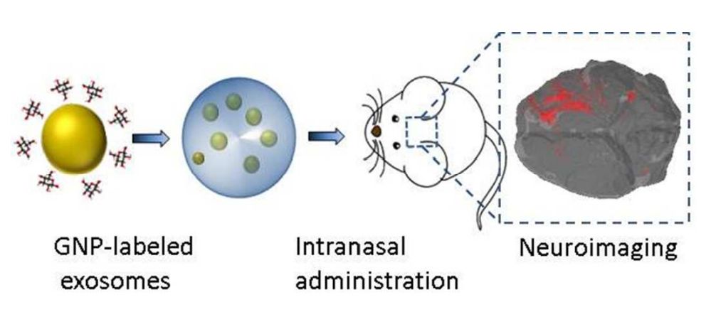
- Applications: CT-based biodistribution; TEM structural confirmation; brain and cardiac delivery studies
- Advantages: Stable CT signal; high electron density; tunable surface chemistry
- Key validation: Loading efficiency; CT attenuation linearity; TEM localization (intra- vs. surface-associated); aggregation status and exosome stability
Quantum Dot (QD) Labeling
- Principle: Semiconductor nanocrystals provide bright, stable fluorescence signals
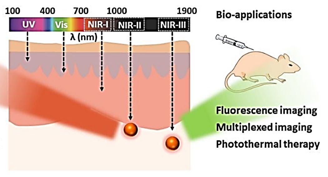
- Applications: Long-term exosome tracking; multiplex uptake studies; live-cell and NIR imaging
- Advantages: High brightness; strong photostability; narrow emission for multiplexing
- Key validation: Conjugation stability; spectral overlap control; aggregation risk; impact on exosome physicochemical properties
Custom / Multimodal Labeling
- Principle: Combination of contrast agents for multi-platform detection
- Applications: Cross-validation studies; multimodal imaging; complex biodistribution questions
- Advantages: Complementary spatial resolution and sensitivity across imaging systems
- Key validation: Cross-agent compatibility; signal interference control; workflow and QC complexity management
Multiple labeling strategies can be integrated or compared within a single project to identify the most suitable approach based on exosome source, imaging platform, and study objective.
Standard Workflow for Magnetic and Contrast Agent Exosome Labeling
- Project Design: Define imaging modality, exosome source, and labeling strategy requirements.
- Sample Evaluation: Assess exosome quality (concentration, size, markers) to ensure labeling suitability.
- Label Selection & Feasibility: Select contrast agent (SPION, GNP, QD, etc.) and confirm loading strategy.
- Labeling Optimization: Optimize conditions to maximize labeling efficiency while preserving exosome integrity.
- Purification & QC: Remove free agents and perform core quality control (purity, integrity, labeling efficiency).
- Imaging Validation & Delivery: Confirm imaging detectability (if required) and deliver QC-verified labeled exosomes with report.
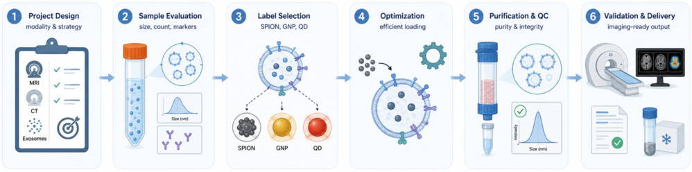 Figure 1. Exosome Magnetic and Contrast Agent Labeling Workflow. (Creative Biostructure)
Quality Control and Characterization
Creative Biostructure applies a targeted QC framework tailored to the labeling agent and downstream imaging needs.
- Particle size & concentration (NTA): Monitors size distribution and particle recovery before and after labeling to detect aggregation or loss during purification.
- Morphology & localization (TEM / Cryo-TEM): Confirms exosome integrity and visualizes nanoparticle incorporation (e.g., intraluminal vs surface-associated loading).
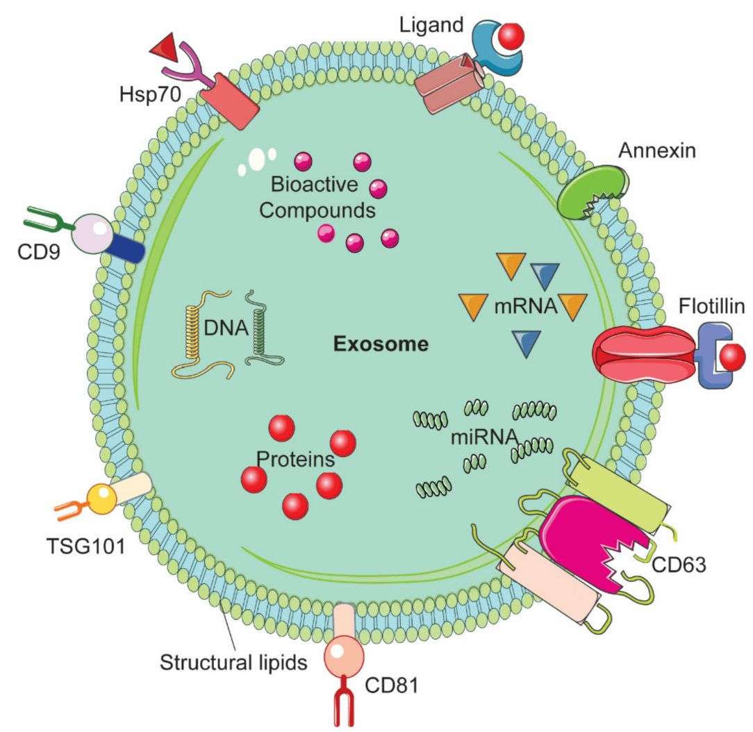
- Marker verification (WB / flow / nano-flow): Confirms preservation of key exosomal markers (CD9, CD63, CD81) after labeling.
- Surface property check (zeta potential): Assesses changes in colloidal stability and surface charge after nanoparticle loading.
- Agent quantification (method-specific): ICP-MS (Fe, Au), relaxometry (R2/R2* for SPION), CT attenuation (GNP), fluorescence readout (QD).
- Free-agent removal validation: Control-based confirmation that unbound nanoparticles are effectively removed post-purification.
- Imaging validation (optional): Phantom or pilot imaging (MRI/CT/fluorescence) to confirm detectability and signal consistency under experimental conditions.
Applications (Research Use Only)
- In vivo exosome biodistribution (MRI/CT): Track organ distribution, clearance, and tissue homing using SPION/USPIO or GNP-labeled exosomes.
- Delivery route comparison: Evaluate how administration routes (IV, intranasal, IP, local injection) affect exosome distribution profiles.
- Targeting efficiency studies: Assess uptake and tissue accumulation of engineered or naturally tropic exosome populations.
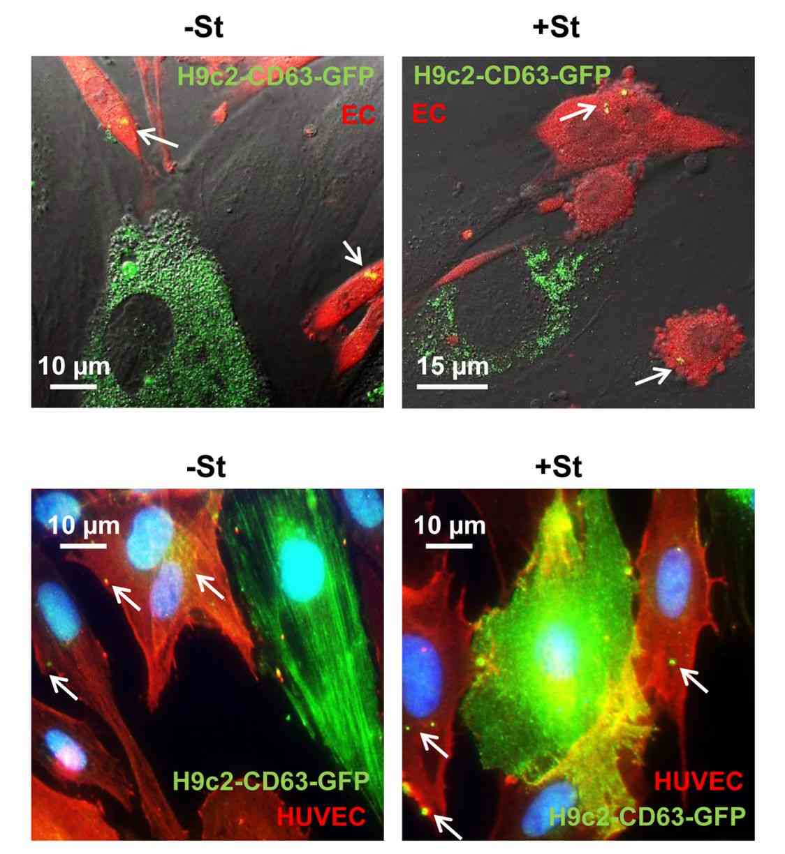
- Cellular uptake & trafficking: Monitor intracellular uptake dynamics using QD-labeled exosomes with fluorescence-based imaging and flow cytometry.
- Multimodal imaging development: Integrate MRI, CT, and optical methods for cross-validated exosome tracking.
- Drug delivery modeling: Use labeled exosomes as imaging surrogates for therapeutic exosome or nanomedicine systems.
What Deliverables Will You Receive
| Deliverable | Details |
|---|---|
| Labeled product | QC-verified labeled exosomes in appropriate buffer, shipped under required storage conditions |
| QC report | Integrated data package: NTA, TEM/cryo-TEM, marker validation, zeta potential, and agent quantification |
| Imaging data | Optional MRI/CT/fluorescence phantom or pilot imaging confirming signal detectability |
| Protocol | Concise workflow summary: labeling strategy, conditions, purification, and handling |
| Purity validation | Free-agent removal verification data |
| Raw data | Instrument raw files available upon request |
| Technical support | Guidance on data interpretation and experimental setup |
How to Get Started
Clients may provide isolated exosomes, conditioned media, or biofluid samples. For raw materials, exosome isolation and pre-characterization services are also available as upstream support.
- Project input: Provide exosome source, imaging modality (MRI, CT, fluorescence, TEM, or multimodal), and research objective.
- Feasibility & proposal: We assess feasibility and define the optimal labeling strategy, workflow, QC plan, and timeline.
Why Choose Creative Biostructure
- Exosome engineering expertise: Hands-on experience across exosome isolation, exosome labeling, and multi-parameter QC for diverse EV sources.
- Multi-platform capability: SPION, GNP, QD, and custom contrast agents supported in one integrated workflow.
- Readout-matched QC: Agent-specific validation (e.g., iron, gold, fluorescence, CT/MRI signal) aligned with imaging modality.
- Artifact control by design: Systematic removal and verification of free nanoparticles in every batch.
- Flexible study design: Adaptable workflows for single-agent, comparative, or multimodal labeling strategies.
- End-to-end service: Integrated isolation, labeling, QC, and imaging-readiness support in one CRO pipeline.
Case Study
Case: MRI Tracking of USPIO-Labeled Stem Cell Exosomes
Background
Non-invasive MRI tracking of exosomes is essential for studying biodistribution and homing. This study established a USPIO-based strategy enabling MRI visualization of stem cell-derived exosomes while preserving vesicle integrity.
Methods
- Labeling strategy: USPIO labeling of parental stem cells prior to exosome isolation
- Agent: Ultrasmall superparamagnetic iron oxide nanoparticles (USPIO, 4-6 nm)
- Imaging: T2/T2*-weighted MRI
- Validation: TEM, Western blot (HSP70/Alix), iron quantification, Prussian blue staining
Results
- Efficient cellular uptake with minimal impact on viability
- Exosomes retained USPIO, confirmed by TEM
- MRI detectable at microgram-level exosome quantities
- In vivo signal confirmed at injection site and validated histologically
Conclusion
USPIO labeling enables robust MRI-based tracking of exosomes while preserving vesicle integrity, supporting preclinical biodistribution and imaging studies.
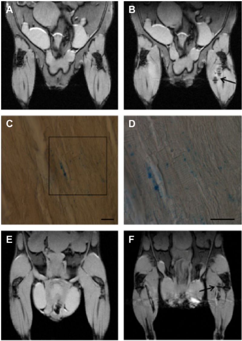 Figure 2. Representative in vivo MRI showing muscle signal changes after intramuscular administration of USPIO-labeled exosomes, with corresponding pre- and post-injection comparison. Histological analysis using Prussian blue staining confirms iron nanoparticle localization at the injection site. (Busato A, et al., 2016)
Ready to track exosome biodistribution with unlimited depth penetration and spatial resolution? Contact us to discuss contrast agent selection, labeling strategy, and imaging protocol design for your study.
References
- Busato A, Bonafede R, Bontempi P, et al. Magnetic resonance imaging of ultrasmall superparamagnetic iron oxide-labeled exosomes from stem cells: a new method to obtain labeled exosomes. International Journal of Nanomedicine. 2016: 2481-2490.
- Busato A, Bonafede R, Bontempi P, et al. Labeling and magnetic resonance imaging of exosomes isolated from adipose stem cells. Current Protocols in Cell Biology. 2017, 75(1): 3.44. 1-3.44. 15.
- Han Z, Liu S, Pei Y, et al. Highly efficient magnetic labelling allows MRI tracking of the homing of stem cell‐derived extracellular vesicles following systemic delivery. Journal of Extracellular Vesicles. 2021, 10(3): e12054.
- Gong L, Weng Y, Zhou W, et al. In vivo CT imaging of gold nanoparticle-labeled exosomes in a myocardial infarction mouse model. Annals of Translational Medicine. 2021, 9(6): 504.
- Ha E, Han Y, Kim M, et al. Quantum Dot‐Based Immunolabelling of Extracellular Vesicles and Detection Using Fluorescence‐Based Nanoparticle Tracking Analysis. Journal of Extracellular Biology. 2025, 4(7): e70072.
Frequently Asked Questions
For any inquiries, our support team is ready to help you get technical support for your research and maximize your experience with Creative Biostructure.