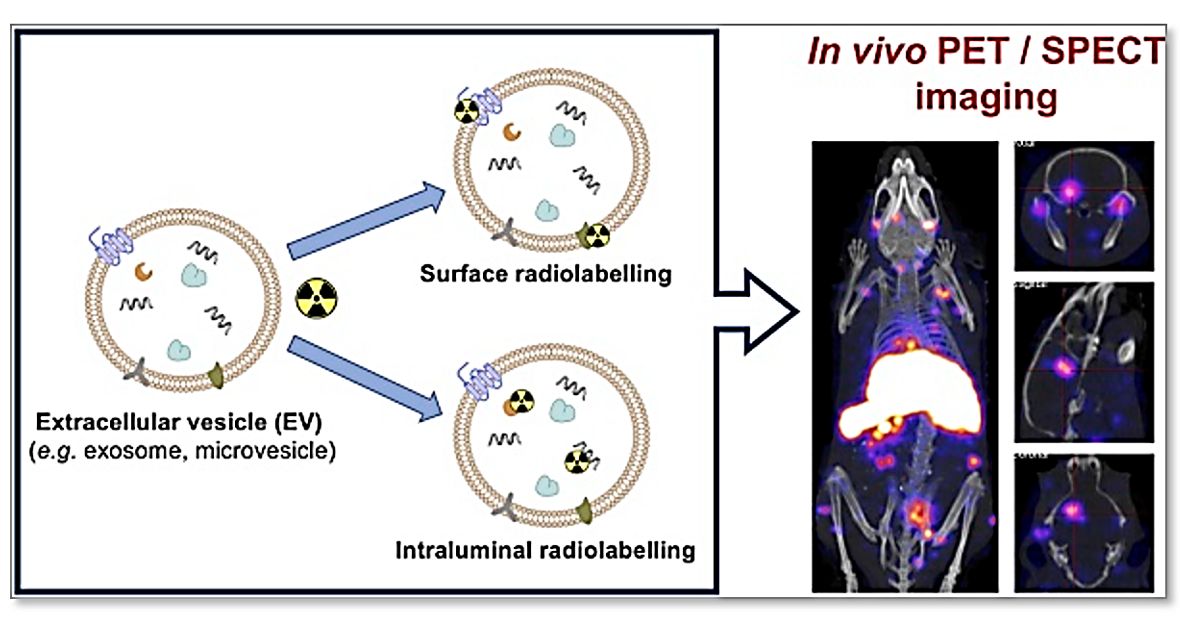
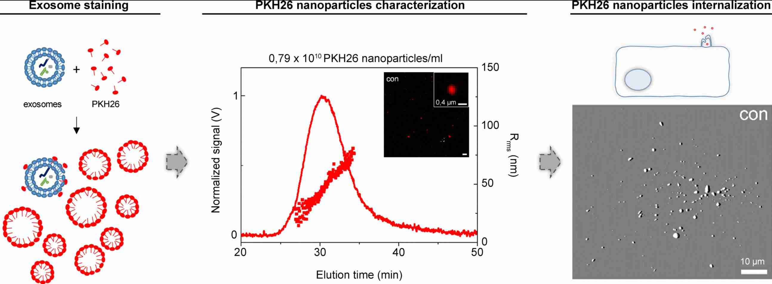
Exosome Fluorescent Labeling Services
Fluorescent labeling is essential for visualizing, tracking, and quantifying exosome behavior in cellular, tissue, and in vivo models. Creative Biostructure provides validated exosome fluorescent labeling services covering lipophilic membrane dyes, surface protein fluorescent conjugation, and exosomal RNA labeling, with optimized protocols designed to preserve exosome integrity, enhance signal-to-noise, and support diverse downstream imaging and analytical workflows.
Why Fluorescent Labeling Matters in Exosome Research
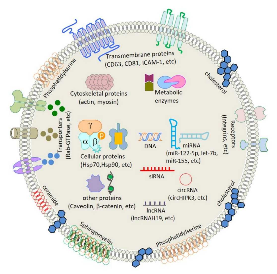
Exosomes are 30-150 nm membrane-bound vesicles released by virtually all cell types, carrying proteins, lipids, and nucleic acids that mediate signaling between cells in both physiological and pathological contexts. Understanding their biodistribution, uptake mechanisms, and functional cargo delivery requires precise, minimally invasive labeling strategies that do not compromise vesicle structure or biological activity.
Fluorescent labeling fulfills these requirements by offering:
- Real-time, live-cell imaging compatibility
- Single-vesicle detection at nanoscale resolution
- Multiplexing capability for co-localization and interaction studies
- Quantitative measurements of cellular uptake and tissue distribution
- Compatibility with standard flow cytometry, confocal microscopy, TIRF, and IVIS imaging platforms
As the field moves toward clinical translation and precision therapeutics, rigorous and reproducible fluorescent labeling workflows are increasingly essential for demonstrating exosome specificity, efficacy, and safety.
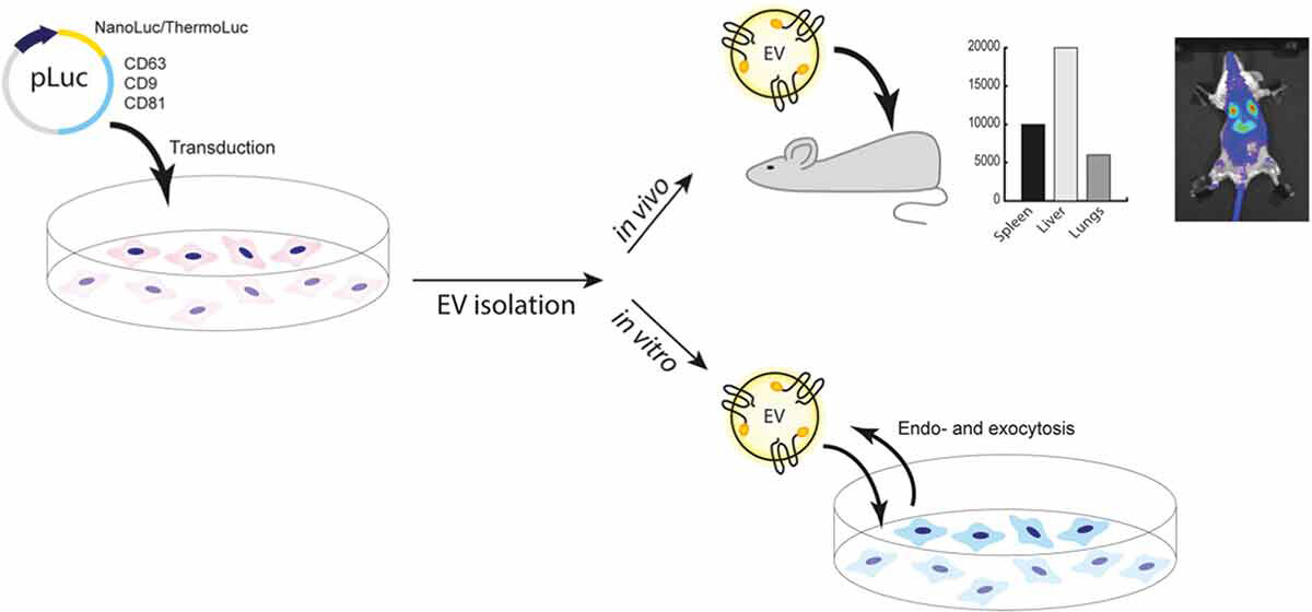
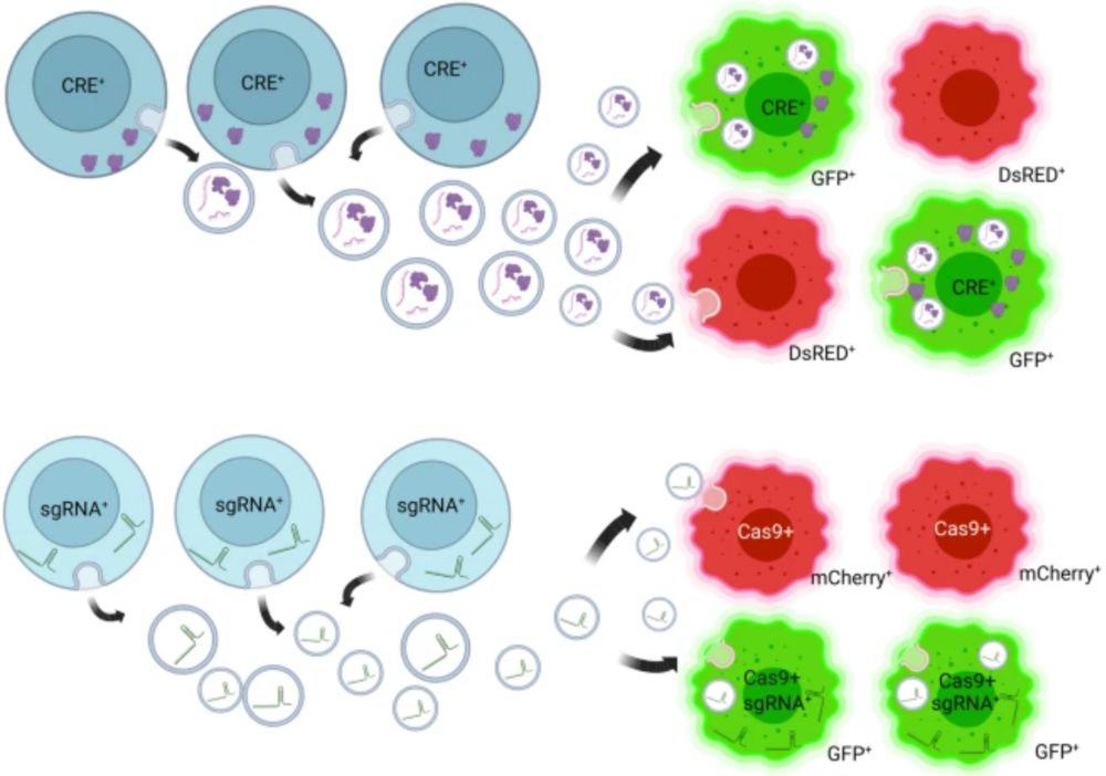 Figure 1. Fluorescence Reporter Strategies for Tracking Functional EV Transfer. (Boudna M, et al., 2024)
Our Fluorescent Labeling Service Portfolio
Creative Biostructure provides three specialized fluorescent labeling services, each designed to address distinct research needs:
1. Exosome Labeling with Lipophilic Dyes (PKH26, DiR, DiI)
Lipophilic carbocyanine and phenoxazine dyes incorporate into the exosomal lipid bilayer, enabling membrane-level fluorescent tracking. PKH26 (red, Ex/Em ~551/567 nm), DiR (near-infrared, Ex/Em ~748/780 nm), and DiI (red-orange, Ex/Em ~549/565 nm) are among the most widely cited dyes in exosome trafficking and biodistribution studies. Our optimized labeling protocols minimize dye aggregation, free dye contamination, and background signal—common pitfalls associated with lipophilic dye labeling.
2. Exosome Surface Protein Fluorescent Labeling Services
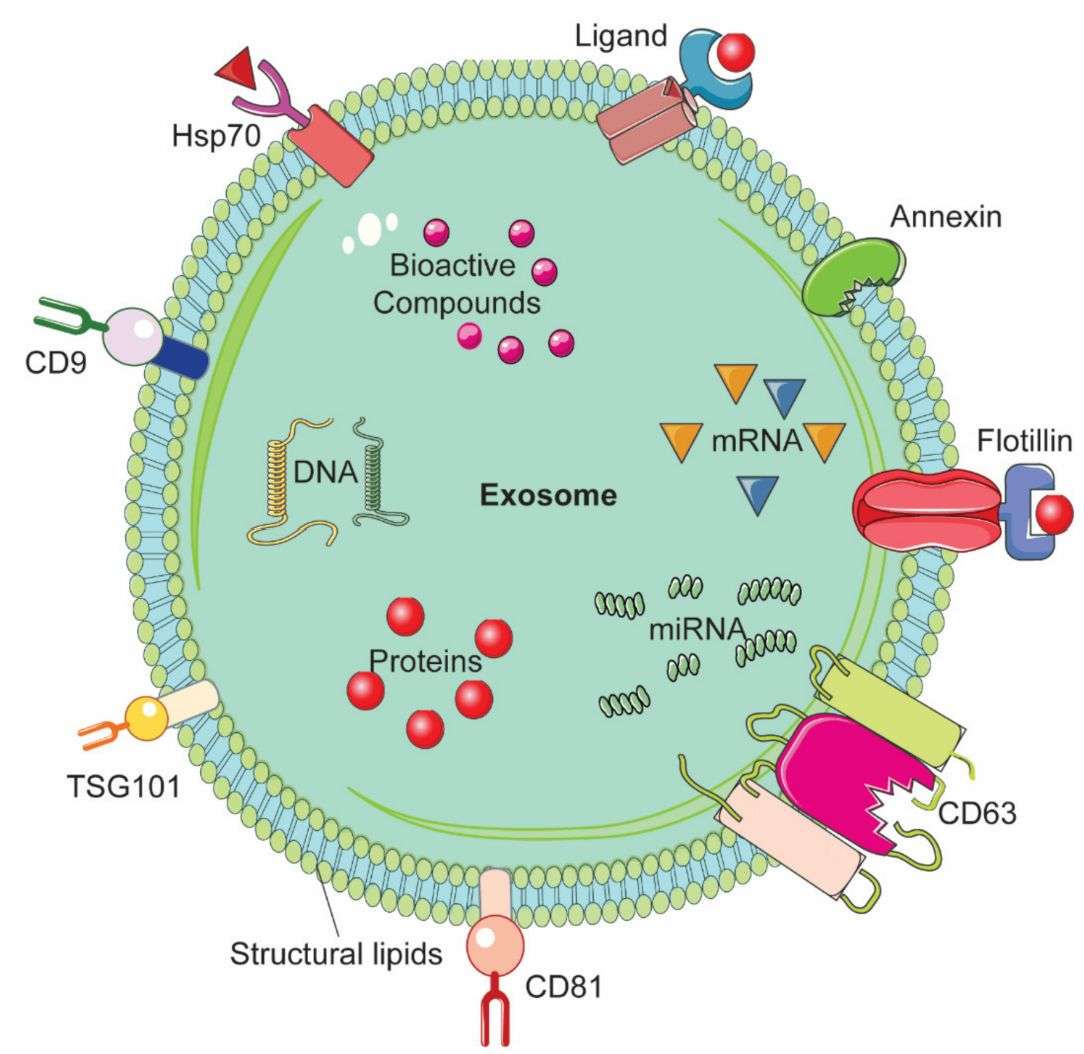
Surface proteins on exosomes, including tetraspanins (CD9, CD63, CD81), EGFR, and integrin family members, can serve as direct labeling targets using fluorophore-conjugated antibodies, NHS-ester reactive dyes, or site-specific chemical strategies. This approach provides mechanistic insights into exosome receptor interactions, uptake routes, and surface proteome dynamics without altering the lipid membrane composition.
3. Exosomal RNA Fluorescent Labeling Services
Exosomal RNAs, including miRNAs, lncRNAs, circRNAs, and mRNAs, are increasingly recognized as functional cargo mediating gene regulatory effects in recipient cells. Our RNA fluorescent labeling services utilize fluorophore-modified oligonucleotide probes, click chemistry-based labeling, and lipophilic dye co-loading strategies to enable direct visualization of RNA cargo delivery and intracellular fate tracking.
Comparison of Fluorescent Labeling Strategies
| Strategy | Target Component | Specificity | Key Applications | Typical Dye/Probe | Special Notes |
|---|---|---|---|---|---|
| Lipophilic dyes (PKH26, DiR, DiI) | Lipid membrane | Membrane-level (all vesicles) | In vivo imaging, uptake assays, biodistribution | PKH26, DiR, DiI, DiO, DiD | Risk of dye aggregates; free dye removal critical by SEC/UC |
| Surface protein labeling | Surface proteins (CD63, CD9, CD81, etc.) | Marker-specific (subpopulation) | Phenotyping, receptor studies, immune tracking | Anti-CD63-FITC, NHS-AF647, NHS-AF488 | Dependent on target protein expression; antibody selection important |
| RNA fluorescent labeling | Lumenal RNA cargo | Cargo-level | RNA delivery, miRNA tracking, intracellular fate | SYTO RNASelect, EU-alkyne, fluorophore-probes | Requires cargo preservation during labeling process |
Standard Workflow
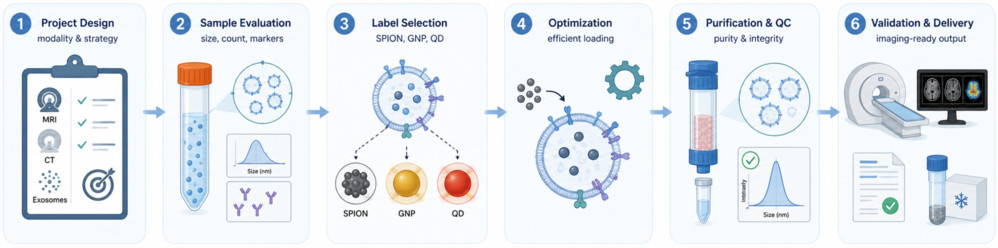
Our workflow ensures efficient exosome fluorescent labeling with reliable quality control and reproducible results.
- Strategy Design: We assess your exosome source, labeling goal, imaging platform, and downstream assays to recommend the optimal labeling method.
- Exosome Preparation: Clients may provide purified exosomes or request exosome production from Creative Biostructure. Samples are checked for size, concentration, purity, and morphology before labeling.
- Fluorescent Labeling: Exosomes are labeled using the selected approach, including lipophilic dyes, surface protein conjugation, or RNA fluorescence labeling.
- Purification: Free dyes, fluorophores, or probes are removed using suitable purification methods to ensure clean labeled exosome preparations.
- QC & Delivery: Labeled exosomes are verified for labeling efficiency, integrity, and downstream usability, then delivered with a data report and technical support.
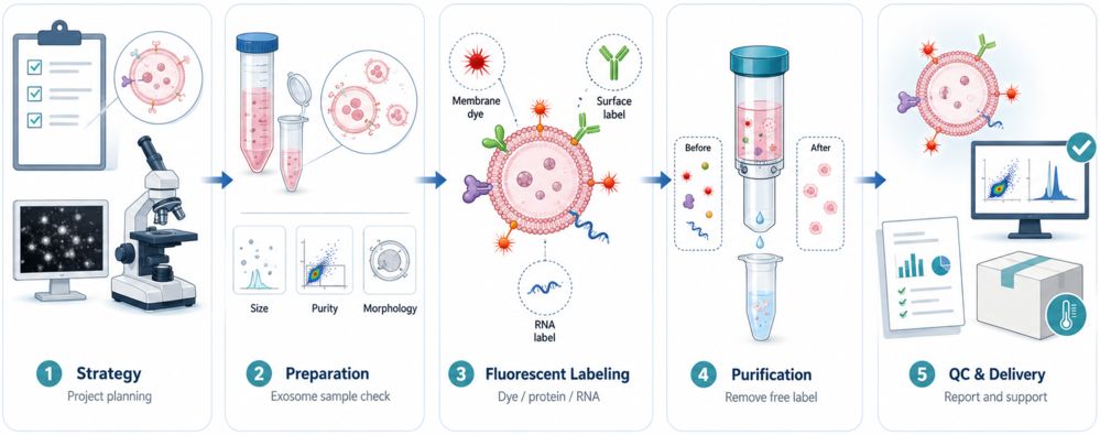 Figure 2. Exosome Fluorescent Labeling Service Workflow. (Creative Biostructure)
Project Initiation Options
| Option | Description | What You Provide | What We Deliver |
|---|---|---|---|
| Customer-Provided Exosomes | You supply purified exosomes; we perform labeling and QC | Purified exosomes (>=1x10^10 particles, characterized) | Labeled exosomes + full QC report |
| Full-Service (End-to-End) | We handle exosome production, labeling, and characterization | Cell line, culture conditions, or project brief | Production records + labeled exosomes + QC report |
| Custom Project | Tailored design for complex or multi-modal requirements | Project consultation required | Custom deliverables per agreement |
Quick Project Kickoff Requirements
To design the optimal labeling strategy, please provide:
- Exosome source (cell line, body fluid, tissue)
- Target labeling modality (membrane, surface protein, or RNA)
- Downstream application (imaging platform, in vitro/in vivo, detection method)
- Preferred fluorophore or dye (if specified)
- Required particle quantity and delivery format
- Timeline and any special handling requirements
What Deliverables Will You Receive
- Fluorescently labeled exosome preparation (specified volume and particle concentration)
- Nanoparticle tracking analysis (NTA) report: size distribution, concentration pre- and post-labeling
- Transmission electron microscopy (TEM) image(s) confirming vesicle morphology
- Labeling efficiency report (fluorescence spectroscopy or flow cytometry data)
- Free-label removal confirmation data (SEC profile or ultracentrifugation documentation)
- Functional uptake assay data (confocal microscopy or flow cytometry, if included)
- Comprehensive technical report with protocols, QC data, and interpretation notes
- Ongoing technical consultation for downstream applications
Characterization and Quality Control
Rigorous quality control is embedded at every stage of our fluorescent labeling workflow. We apply a multi-parameter validation strategy to confirm that labeled exosomes meet defined quality benchmarks before delivery.
Standard QC Parameters:
- Particle size and size distribution: NTA and DLS
- Particle morphology: TEM imaging
- Fluorescence signal intensity and spectral profile: Fluorescence spectrometer
- Labeling efficiency: Flow cytometry (NanoFCM for single-vesicle analysis where applicable)
- Free dye/probe removal confirmation: SEC, FCS, or fluorescence quenching assays
- Vesicle integrity: Zeta potential, Cryo-EM (on request)
- Exosome marker validation: Western blot for CD63, CD9, CD81, HSP70 (on request)
- Functional uptake verification: Confocal microscopy with recipient cells
Applications
Creative Biostructure's fluorescently labeled exosomes support a diverse range of preclinical and discovery research applications:
- Exosome biodistribution and organ tropism mapping in mouse models
- Cellular uptake, endocytosis pathway, and intracellular trafficking studies
- Tumor microenvironment communication and cancer cell targeting research
- Exosome-mediated drug delivery and cargo release studies
- Immune cell activation and immunomodulation studies
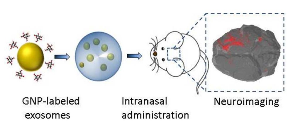
- Blood-brain barrier (BBB) crossing and neurological targeting research
- Exosome pharmacokinetics and half-life studies in preclinical models
- Co-localization and fusion studies with recipient cell organelles
- Biomarker discovery and diagnostic assay development
Why Choose Creative Biostructure
- Optimized labeling workflows: Protocols are tailored to each exosome source, fluorophore type, and downstream application.
- Stringent free-label removal: Dedicated purification steps help reduce background signals and improve data reliability.
- Comprehensive QC: Labeled exosomes are assessed for size, morphology, labeling efficiency, integrity, and functional readiness.
- Flexible service options: Support customer-provided samples or full workflows from exosome production to labeled sample delivery.
- Application-oriented support: Scalable workflows for in vitro tracking, biodistribution studies, and preclinical research.
Case Study
Case: Live Tracking of Endogenous Exosomes in Zebrafish
Background
Real-time visualization of endogenous exosomes in vivo remains challenging due to their small size and dynamic behavior. This study established a zebrafish model to track exosome release, circulation, uptake, and functional impact using fluorescence-based labeling.
Methods
- Labeling strategy: CD63-pHluorin fluorescent reporter
- Model: Transparent zebrafish embryos
- Tracking focus: Exosome biogenesis, blood circulation, cellular uptake, and tissue distribution
- Analysis: Live fluorescence imaging, electron microscopy, NTA, immunogold labeling, and proteomics
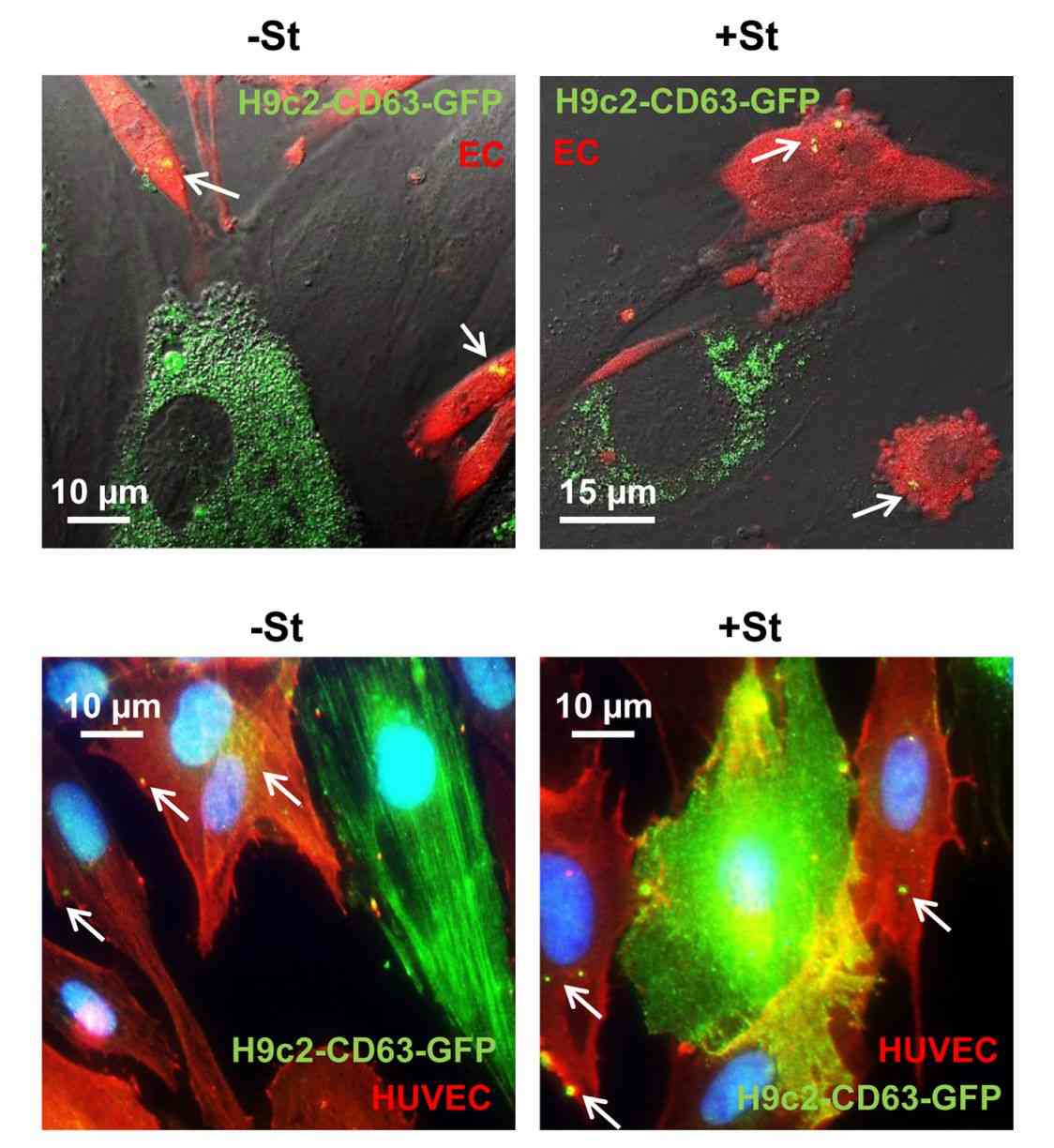
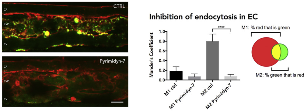 Figure 3. Dynamin-Dependent Uptake of Fluorescently Labeled Exosomes. (Verweij F J, et al., 2019)
Results
- Real-time tracking: Individual CD63-positive exosomes were visualized in vivo.
- Clear biodistribution: Labeled exosomes entered the bloodstream and accumulated in specific vascular regions.
- Target cell uptake: Exosomes were internalized by endothelial cells and macrophages.
- Mechanistic insight: Uptake was associated with dynamin-dependent endocytosis and scavenger receptor pathways.
- Functional relevance: Reduced exosome release affected caudal vein plexus development, suggesting trophic support functions.
Conclusion
Fluorescence-based exosome labeling enables high-resolution in vivo tracking of exosome release, biodistribution, cellular uptake, and biological function.
Looking to visualize exosome uptake, biodistribution, or cargo transfer with greater confidence? Creative Biostructure provides tailored exosome labeling strategies based on your exosome source, labeling target, imaging platform, and research goals. Contact us to start your project with a validated exosome fluorescent labeling solution.
References
- Verweij F J, Revenu C, Arras G, et al. Live tracking of inter-organ communication by endogenous exosomes in vivo. Developmental Cell. 2019, 48(4): 573-589. e4.
- Boudna M, Campos A D, Vychytilova-Faltejskova P, et al. Strategies for labelling of exogenous and endogenous extracellular vesicles and their application for in vitro and in vivo functional studies. Cell Communication and Signaling. 2024, 22(1): 171.
Frequently Asked Questions
For any inquiries, our support team is ready to help you get technical support for your research and maximize your experience with Creative Biostructure.