Exosome Surface Modification Service with Aptamers
Aptamers—short single-stranded DNA or RNA oligonucleotides selected through the SELEX (Systematic Evolution of Ligands by Exponential Enrichment) process—offer exceptional binding affinity and specificity toward a wide range of molecular targets. When conjugated to the exosome surface, aptamers transform native extracellular vesicles into precision-guided nanocarriers capable of receptor-targeted recognition, selective cellular uptake, and controlled cargo delivery.
At Creative Biostructure, we provide customized exosome surface modification service with aptamers, enabling researchers to engineer functionally enhanced exosomes for targeted delivery studies, biosensing applications, and preclinical therapeutic investigations. Our platform integrates aptamer design, validated conjugation chemistries, and rigorous characterization to ensure optimal biological performance.
Why Aptamer-Based Exosome Surface Modification
Although exosomes exhibit intrinsic biocompatibility and natural cellular communication capabilities, their native surface profiles often lack the targeting precision required for selective cell interaction. Aptamer functionalization addresses this limitation by providing programmable, high-affinity recognition elements that can be customized to virtually any surface antigen or receptor.
Key advantages of aptamer-modified exosomes include:
- High binding affinity and specificity: Dissociation constants in the nanomolar to picomolar range against defined molecular targets
- Low immunogenicity: Oligonucleotide-based ligands with minimal immune activation risk compared to antibodies
- Chemical versatility: Amenable to diverse conjugation chemistries including cholesterol insertion, click chemistry, and biotin-streptavidin coupling
- Customizable by design: Sequences engineered or selected to target specific biomarkers such as nucleolin, EpCAM, EGFR, PSMA, and MUC1
- Scalable synthesis: Chemical synthesis enables high-purity, batch-consistent aptamer production without biological manufacturing
- Stability: Chemical modifications (e.g., phosphorothioate backbones, 2′-F/OMe substitutions) enhance nuclease resistance
These properties make aptamer-based surface modification a compelling alternative and complement to antibody or peptide functionalization strategies in advanced exosome engineering.
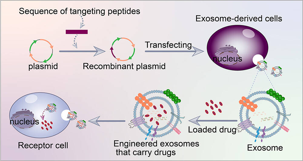
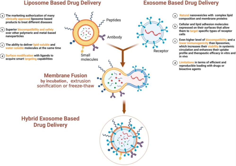
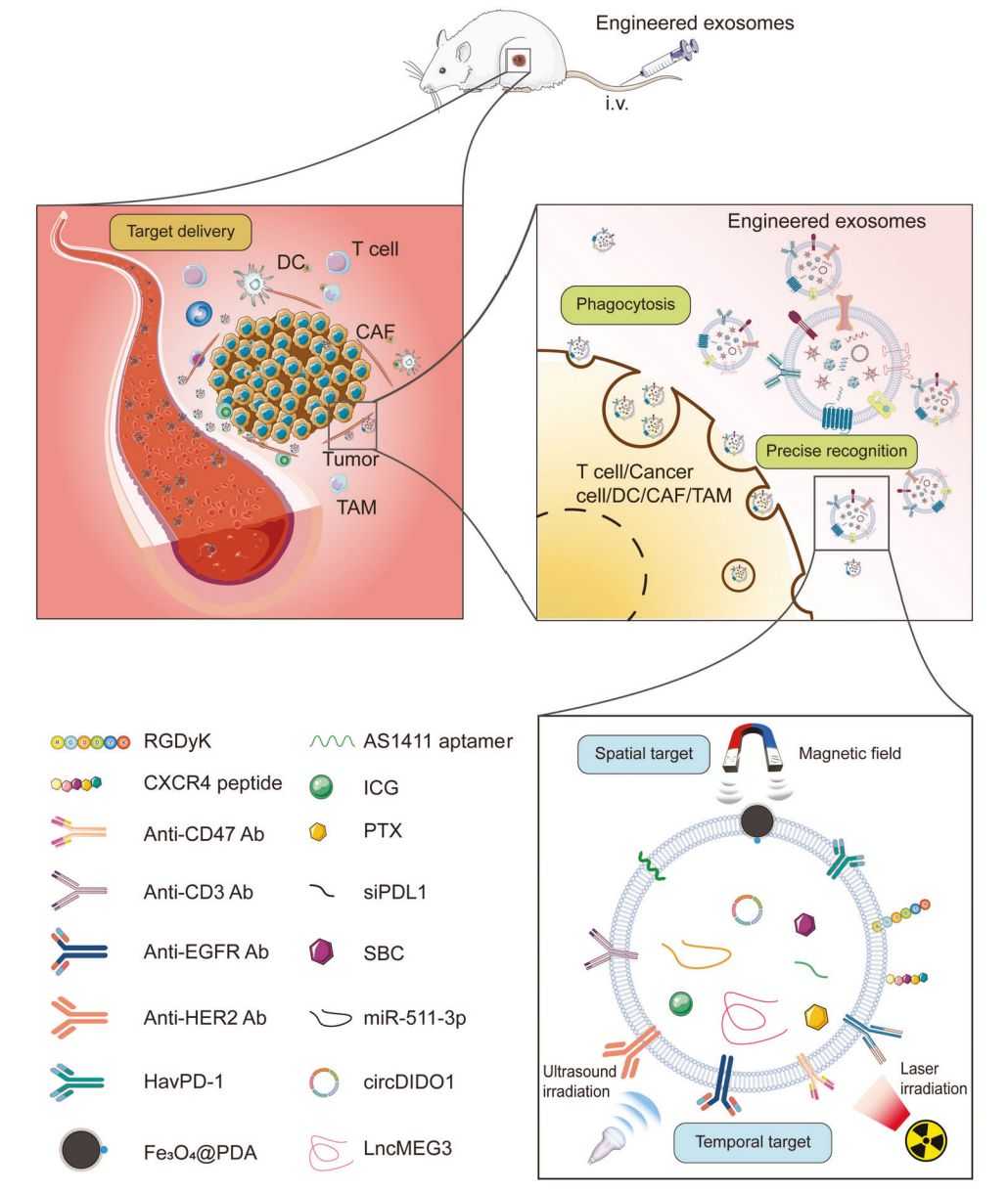
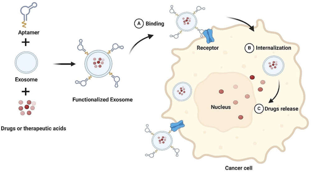 Figure 1. Aptamer-Functionalized Exosome for Targeted Cancer Therapy. (Wu Y, et al., 2024)
Comparison of Exosome Surface Modification Strategies
Aptamer-based modification occupies a unique positioning among exosome engineering strategies:
| Feature | Aptamers | Antibodies | Peptides & Ligands |
|---|---|---|---|
| Molecular Size | Small (6-30 kDa) | Large (~150 kDa) | Small (<5 kDa) |
| Target Specificity | High (picomolar-nanomolar Kd) | High | Moderate-High |
| Immunogenicity | Very Low | Moderate | Low |
| Tissue Penetration | Good | Limited | Excellent |
| Stability | High (with modification) | Moderate | High |
| Custom Selectability | Yes (SELEX) | Limited | Limited |
Our Aptamer Functionalization Strategies
We offer multiple validated aptamer conjugation and display platforms, each optimized for exosome integrity, functionalization efficiency, and downstream performance. Strategy selection is guided by aptamer type, target biology, and application requirements.
1. Cholesterol- or Lipid-Anchored Aptamer Insertion (Post-Insertion Method)
Aptamers conjugated with hydrophobic anchors (e.g., cholesterol, DSPE lipid tails) spontaneously intercalate into the exosome lipid bilayer under mild conditions. This approach preserves vesicle structure and biological activity while enabling efficient surface display.
- Cholesterol-DNA/RNA aptamer post-insertion
- DSPE-PEG-aptamer conjugate integration
- Mild, aqueous-phase incubation protocol
Best for: Preserving exosome membrane integrity with controlled surface density
2. Covalent Chemical Conjugation
Aptamers are covalently attached to exosome membrane proteins or modified lipid head groups through well-characterized bioconjugation reactions, providing stable, long-lasting surface modification resistant to physiological conditions.
- Click chemistry (azide–alkyne, DBCO–azide cycloaddition)
- NHS-ester / EDC coupling to amine groups
- Maleimide–thiol conjugation for site-specific attachment
Best for: Stable and reproducible covalent surface modification
3. Biotin-Streptavidin Bridge Coupling
Biotinylated aptamers are anchored to streptavidin pre-conjugated onto exosome surfaces via NHS-ester chemistry, enabling modular, high-affinity aptamer display. This strategy is widely used in biosensing and detection research.
- Pre-functionalization of exosomes with streptavidin
- High-affinity biotin–streptavidin binding (Kd ~10⁻¹⁵ M)
- Supports multiplex aptamer display
Best for: Flexible, modular aptamer loading for detection and capture applications
4. RNA Nanotechnology-Based Display
Using RNA nanostructure design, aptamers are incorporated into engineered RNA scaffolds (e.g., arrowhead nanoparticles) that anchor to the exosome outer membrane via cholesterol or lipid tails, presenting aptamers in optimal orientations for target engagement.
- Programmable RNA scaffold design with embedded aptamer modules
- Precise control of aptamer orientation and density
- Compatible with multivalent aptamer presentation
Best for: Structural precision and multivalent display studies
5. Hybrid Functionalization Strategies
Aptamer surface display can be combined with other exosome engineering approaches—including PEGylation, cargo loading, and membrane fusion—to create multifunctional exosome systems with tunable properties.
- Aptamer + PEGylation for stealth and targeted delivery combination
- Aptamer + siRNA/miRNA cargo co-loading
- Dual aptamer display for multi-receptor targeting
Best for: Advanced research requiring simultaneous targeting, stealth, and cargo delivery
Representative Aptamers Supported
| Aptamer | Target | Application Area |
|---|---|---|
| Sgc8 / TD05 | PTK7 / CCRF-CEM (leukemia) | Cancer cell-selective uptake |
| MUC1 aptamer | MUC1 (breast, ovarian cancer) | Targeted oncology research |
| EpCAM aptamer | EpCAM (epithelial cancers) | CTC detection, targeted delivery |
| PSMA aptamer | PSMA (prostate cancer) | Prostate cancer targeting |
| CL4 aptamer | EGFR | NSCLC, TNBC research |
| hBS01 | Transferrin receptor (BBB transport) | CNS drug delivery research |
| CD63 aptamer | Exosome surface protein CD63 | Exosome capture, EAA loading |
| Custom / SELEX-derived | Client-defined target | Any receptor or biomarker |
Service Workflow
Consultation & Target Assessment
Evaluate receptor biology, aptamer options, and project goals, with feasibility and experimental design recommendations.
Aptamer Selection & Validation
Select literature-validated aptamers or initiate custom SELEX, confirming binding affinity.
Conjugation Strategy Design
Optimize surface modification based on aptamer structure, exosome source, and application.
Exosome Production or Sample Assessment
Produce exosomes from cell lines or assess client samples for quality and compatibility.
Aptamer Surface Modification & Purification
Execute conjugation under optimized conditions, removing unconjugated aptamers and byproducts.
Characterization & Delivery
Verify modified exosomes and provide delivery with full documentation and technical support.
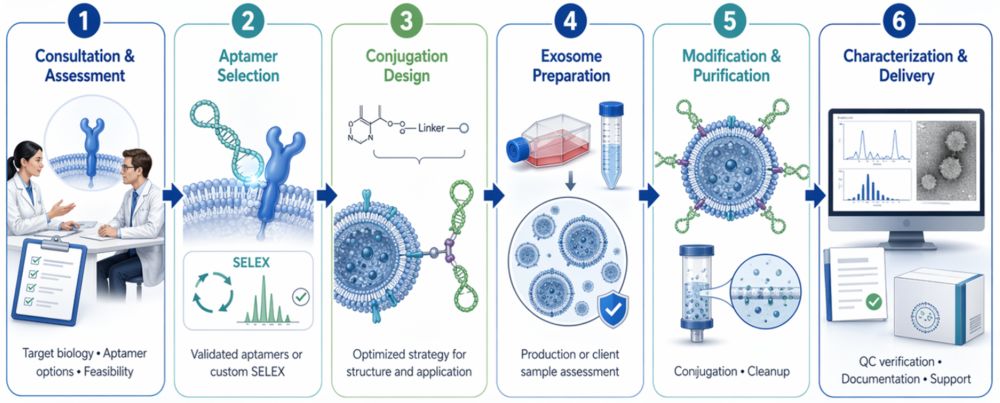 Figure 2. Workflow of Exosome Surface Modification Service with Aptamers. (Creative Biostructure)
Characterization and Quality Control
We provide multi-dimensional analytical validation to confirm successful aptamer surface modification and maintain exosome integrity:
- Particle size and size distribution: NTA (nanoparticle tracking analysis) and DLS (dynamic light scattering)
- Morphology analysis: TEM (transmission electron microscopy) or Cryo-EM
- Zeta potential measurement: Surface charge assessment before and after modification
- Aptamer surface display confirmation: Fluorescence-labeled aptamer tracking, flow cytometry
- Aptamer copy number quantification: Fluorescence quantification or mass spectrometry-based analysis
- Binding affinity verification: EMSA, SPR, or cell-based binding assays with target-positive and target-negative controls
- Cellular uptake and internalization studies: Confocal microscopy, flow cytometry with relevant cell models
- Exosome protein marker validation: Western blot (CD9, CD63, CD81, TSG101) to confirm vesicle identity
- Stability testing: Colloidal stability in physiological buffers; nuclease resistance testing (where applicable)
Applications of Aptamer-Modified Exosomes
Aptamer-functionalized exosomes support a broad spectrum of research applications where high targeting precision and programmable surface chemistry are essential:
| Application Area | Description |
|---|---|
| Targeted Cancer Therapy Research | Receptor-specific delivery of chemotherapeutics, siRNA, or miRNA to tumor cells expressing EpCAM, MUC1, EGFR, or PSMA |
| Blood-Brain Barrier (BBB) Transport Studies | Aptamer-guided exosome transcytosis across the BBB using transferrin receptor-targeting aptamers for CNS drug delivery research |
| Nucleic Acid Therapeutics Delivery | Loading and targeted delivery of siRNA, antisense oligonucleotides (ASO), PMO, and miRNA via aptamer-cargo co-loading strategies |
| Liquid Biopsy & Biosensing | Aptamer-functionalized exosomes or surfaces for capture and detection of disease-specific exosomes from clinical samples (serum, plasma, urine) |
| Immunotherapy Support | Aptamer-guided delivery of immune-modulating payloads; investigation of aptamer-exosome platforms in checkpoint inhibitor combination strategies |
| Cell-Type Selective Interaction Studies | Investigation of receptor-mediated endocytosis and intracellular trafficking in defined cell populations |
How to Start Your Project
We provide flexible project initiation options to accommodate different research scenarios. You may choose to provide your own exosomes and/or aptamers, or engage our end-to-end service.
Project Initiation Options
| Option | Description | Best For | What You Need to Provide |
|---|---|---|---|
| Client-Provided Exosomes & Aptamer | We perform surface modification using your supplied exosome samples and aptamer sequences or molecules. | Researchers with established exosome systems or proprietary aptamers |
|
| Client-Provided Aptamer Only | We produce exosomes in-house and execute aptamer surface modification per your specifications. | Researchers with characterized aptamers but without dedicated exosome production |
|
| End-to-End Service (Recommended) | Full workflow: exosome production, aptamer selection or sourcing, surface modification, and comprehensive validation. | Researchers seeking a turnkey solution with minimal experimental burden |
|
Quick Project Kickoff Requirements
| Information Type | Details |
|---|---|
| Target Information | Target receptor, cell type, or disease model of interest |
| Aptamer Information | Known aptamer (if available) or target description for selection/recommendation |
| Application Goal | Targeted uptake, cargo delivery, biosensing, BBB research, etc. |
| Exosome Source | Cell line of origin, isolation method, concentration (if client-provided) |
| Special Requirements | Specific conjugation chemistry preference, QC assays, or downstream use conditions |
What Deliverables Will You Receive
Our service provides clearly defined, research-ready deliverables to support reproducibility, validation, and downstream applications:
| Category | Description |
|---|---|
| Aptamer-Modified Exosome Samples | Surface-functionalized exosomes with defined aptamer type, particle concentration, volume, and storage conditions |
| Aptamer Surface Display Confirmation | Fluorescence-based tracking or biochemical assay data confirming aptamer conjugation and surface density |
| Physicochemical Characterization | Size distribution (NTA/DLS), morphology (TEM), zeta potential, and purity assessment |
| Functional Evaluation (Optional) | Comparative cellular uptake analysis of aptamer-modified vs. unmodified exosomes in target cell models |
| Methods & Protocol Summary | Detailed conjugation strategy, key experimental parameters, and aptamer source/sequence information |
| Project Report | Consolidated results, data interpretation, QC assessment, and technical insights |
| Technical Support | Post-delivery consultation for downstream applications, troubleshooting, and experimental guidance |
Why Choose Creative Biostructure
- Expertise in exosome engineering with validated modification platforms
- Extensive aptamer library, including Sgc8, EpCAM, PSMA, MUC1, and custom sequences
- Multiple conjugation strategies tailored to project needs
- Rigorous quality control ensuring vesicle integrity
- Flexible project models: partial or full turnkey solutions
- Scalable workflows for exploratory and preclinical research
Case Study
Case: Aptamer-Functionalized Exosomes for Targeted Doxorubicin Delivery in Colorectal Cancer
Background
Doxorubicin (DOX) suffers from poor targeting and severe side effects. This study developed aptamer-functionalized exosomes (DOX-Apt-Exo) for targeted delivery to nucleolin-expressing colorectal cancer cells.
Methods
HEK-293-derived exosomes were loaded with DOX and functionalized with aptamer using EDC/NHS chemistry. Characterization was done via DLS, TEM, and flow cytometry.
Results
- In vitro: DOX-Apt-Exo showed enhanced uptake and lower cytotoxicity compared to non-targeted exosomes.
- In vivo: DOX-Apt-Exo reduced tumor size by 65%, significantly outperforming free DOX (72%) and non-targeted DOX-Exo.
- Biodistribution: Exosomes accumulated at tumor sites, with minimal uptake in non-target tissues.
Conclusion
Aptamer-functionalized exosomes enhance DOX delivery and efficacy, providing a safe and effective solution for targeted cancer therapy.
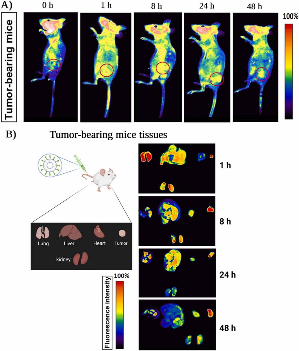 Figure 3. In Vivo Biodistribution of FAM-Labeled Aptamer-Functionalized Exosomes in Tumor-Bearing Mice. (Hosseini N F, et al., 2022)
Design aptamer-functionalized exosomes tailored to your specific research targets and delivery objectives. Our team will develop a custom engineering strategy optimized for your application, from aptamer selection to functional validation. Contact us to discuss your project and receive a personalized proposal.
References
- Hosseini N F, Amini R, Ramezani M, et al. AS1411 aptamer-functionalized exosomes in the targeted delivery of doxorubicin in fighting colorectal cancer. Biomedicine & Pharmacotherapy. 2022, 155: 113690.
- Han G, Zhang Y, Zhong L, et al. Generalizable anchor aptamer strategy for loading nucleic acid therapeutics on exosomes. EMBO Molecular Medicine. 2024, 16(4): 1027.
- Wu Y, Cao Y, Chen L, et al. Role of exosomes in cancer and aptamer-modified exosomes as a promising platform for cancer targeted therapy. Biological Procedures Online. 2024, 26(1): 15.
Frequently Asked Questions
For any inquiries, our support team is ready to help you get technical support for your research and maximize your experience with Creative Biostructure.