Exosome Labeling Services
Creative Biostructure provides comprehensive exosome labeling services to support biodistribution tracking, imaging, and functional studies in vitro and in vivo. Our solutions enable reliable detection of extracellular vesicles (EVs) across fluorescence microscopy, bioluminescence imaging, radiolabeling-based tracking, MRI/CT contrast imaging, genetic reporter systems, and biotin-mediated downstream applications. Built on validated protocols, rigorous quality control, and deep expertise in EV biology, we deliver precisely labeled exosome preparations tailored to diverse research and industrial needs.
What Is Exosome Labeling
Exosome labeling is the process of introducing detectable markers—such as fluorescent dyes, luminescent enzymes, radioisotopes, magnetic or contrast agents, reporter constructs, or biotin groups—into or onto exosomes for reliable detection. Since native exosomes are nanoscale vesicles with limited intrinsic signal, labeling enables tracking of their biodistribution, cellular uptake, subcellular localization, and pharmacokinetic behavior in complex biological systems.
Common labeling strategies include:
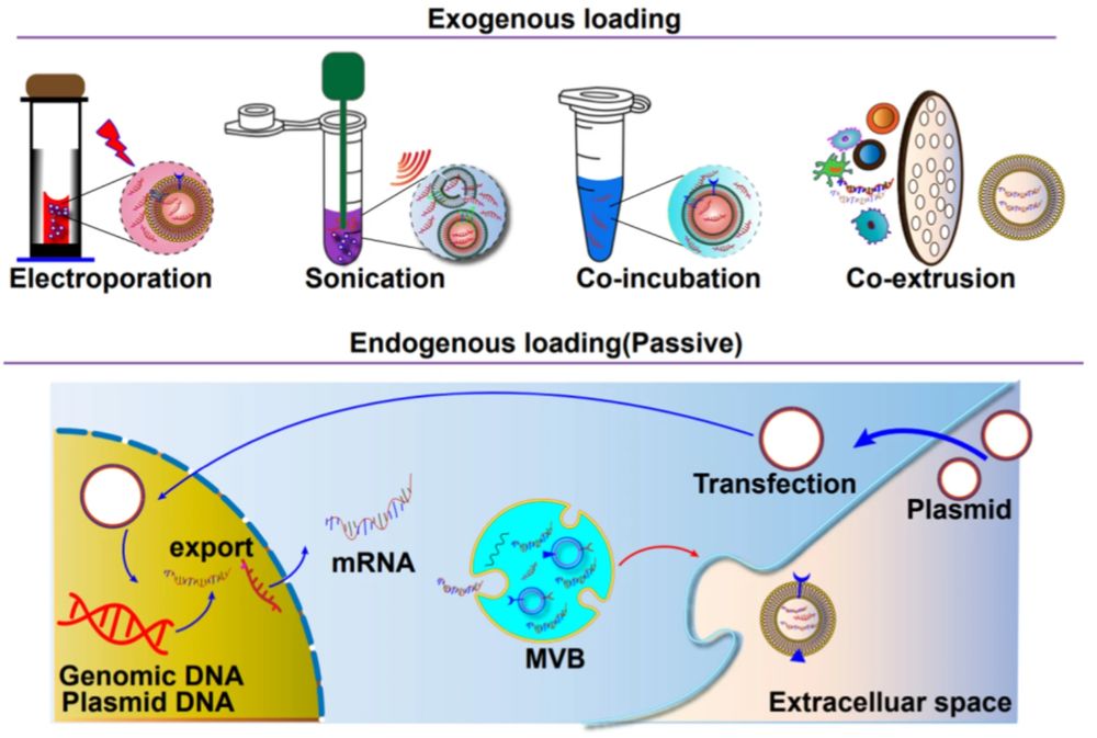
- Exogenous labeling: Post-isolation conjugation of dyes, tracers, magnetic agents, or biotin to purified exosomes.
- Endogenous labeling: Engineering producer cells to express reporter proteins, such as GFP, or luciferase, fused with exosome-associated proteins.
The optimal strategy depends on the detection platform, sensitivity requirements, experimental model, and intended in vitro or in vivo application.
Why Is Proper Exosome Labeling Critical
Poorly controlled labeling can alter exosome surface chemistry, impair biological function, or generate signal artifacts from free dye or unbound reagents. Rigorous labeling and purification are therefore essential for:
- Accurate biodistribution and pharmacokinetic studies
- Reliable cellular uptake and trafficking experiments
- High-resolution imaging without background interference
- Quantitative cargo tracking in therapeutic development pipelines
- Multi-modal imaging combining two or more detection strategies
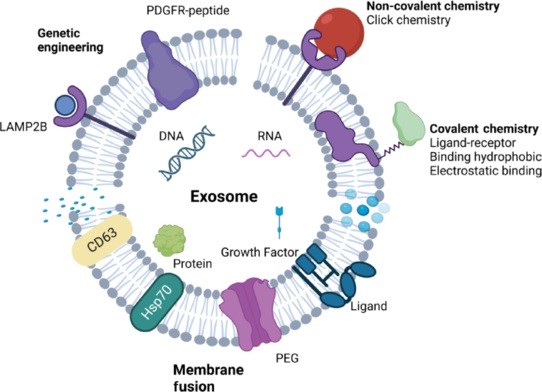
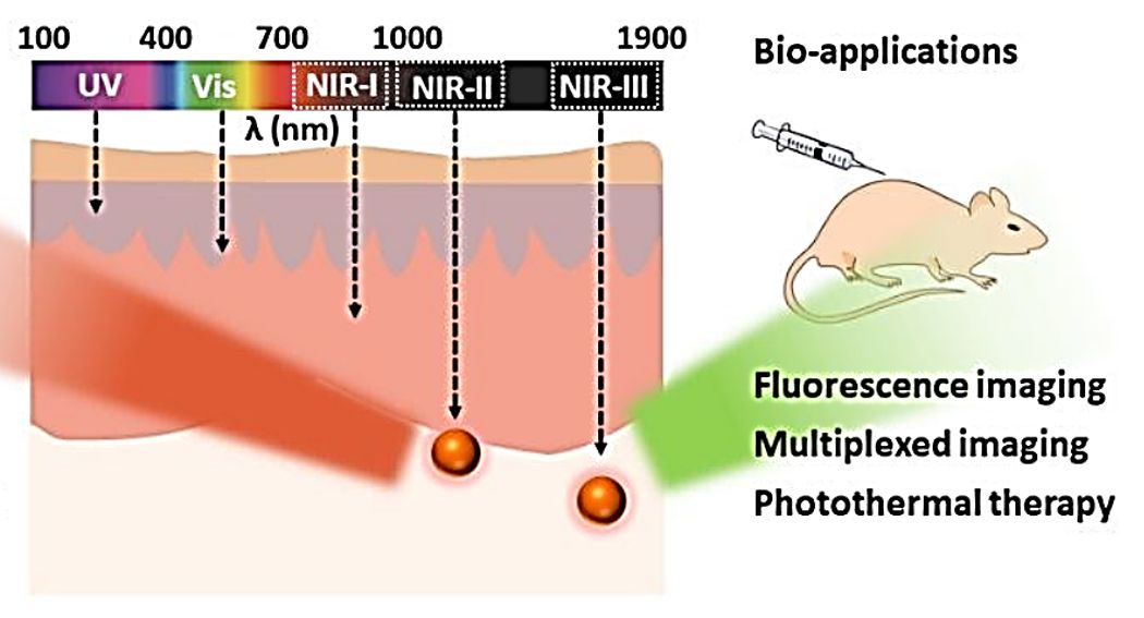
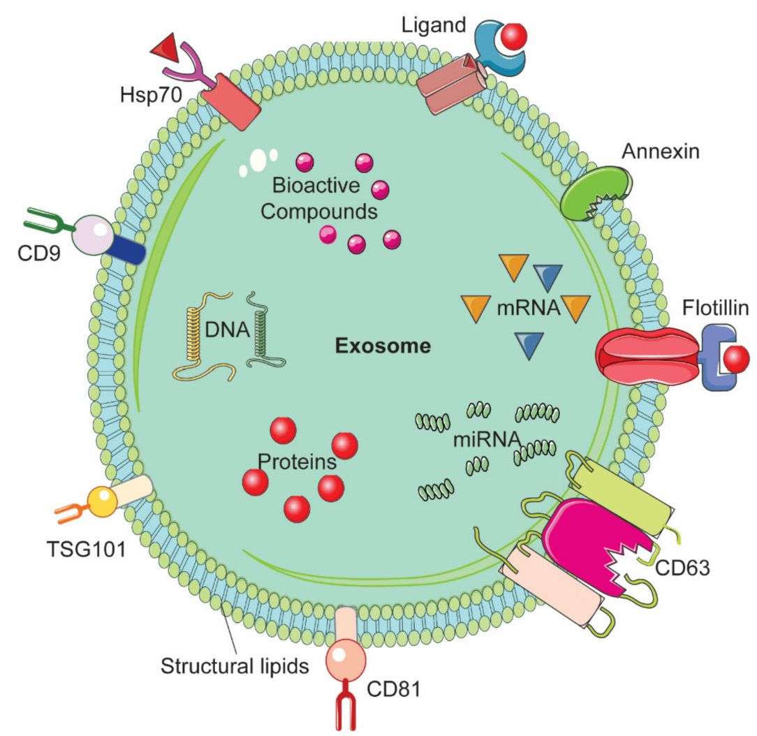
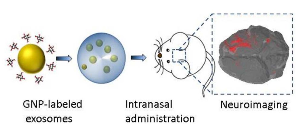
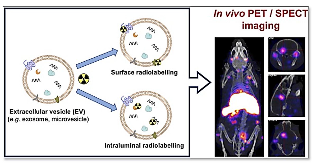
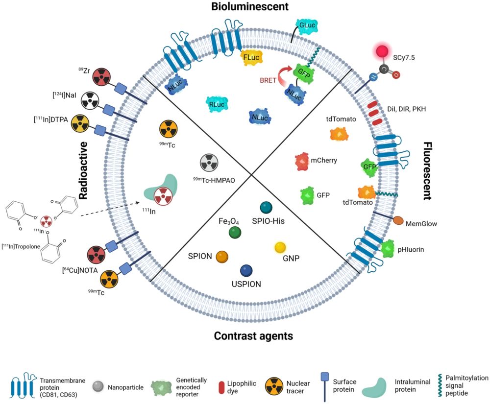 Figure 1. Exosome Imaging and Labeling Strategies. (Boudna M, et al., 2024)
Our Exosome Labeling Capabilities
Creative Biostructure provides comprehensive exosome labeling services matched to specific imaging modalities and research goals. For each project, we optimize label selection, reagent concentration, incubation conditions, purification, and quality control to achieve specific signals while preserving exosome integrity.
| Service | Key Features | Primary Applications |
|---|---|---|
| Exosome Fluorescent Labeling | Membrane, surface, and intraluminal labeling using lipophilic dyes, amine-reactive dyes, antibody–fluorophore conjugates, and multicolor strategies | Confocal and TIRF microscopy, flow cytometry, cellular uptake, co-localization, and single-EV analysis |
| Exosome Bioluminescent Labeling | Luciferase reporters fused to exosome-enriched proteins such as CD63, CD9, and CD81 | Sensitive longitudinal BLI, in vivo biodistribution, clearance, and tissue uptake studies |
| Exosome Radiolabeling | Direct or chelator-mediated labeling with radionuclides such as 125I, 99mTc, 64Cu, and 18F, with radiochemical purification and QC | Quantitative PET/SPECT imaging, pharmacokinetics, organ distribution, and whole-body tracking |
| Exosome Magnetic and Contrast Agent Labeling | SPION/USPIO loading, gold nanoparticle labeling, and custom or multimodal contrast-agent strategies | MRI and CT tracking, deep-tissue biodistribution, anatomical localization, and multimodal imaging validation |
Standard Workflow
Our standardized workflow ensures consistent execution, rigorous quality control, and transparent project delivery:
- Project Consultation & Strategy Design: Define detection modality, sample type, study objectives, and the most suitable labeling approach.
- Exosome Preparation & Initial Assessment: Isolate exosomes from specified sources or evaluate client-provided materials using methods such as NTA and Western blot.
- Labeling Optimization: Select labeling reagents, conjugation chemistry, reagent concentration, and reaction conditions through small-scale feasibility testing.
- Scale-Up Labeling Execution: Perform controlled and reproducible labeling reactions at the target project scale.
- Purification & Quality Control: Remove free dyes or unconjugated reagents and characterize labeled exosomes for purity, size, marker expression, and labeling efficiency.
- Delivery & Technical Support: Provide labeled exosome preparations with documentation, data summary, and follow-up scientific consultation.
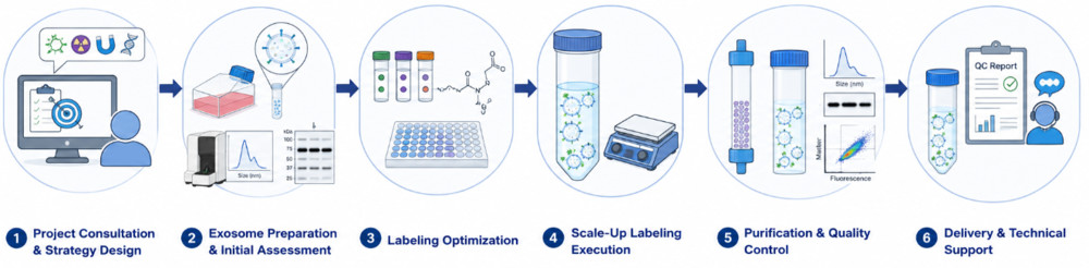 Figure 2. Project Workflow for Exosome Labeling. (Creative Biostructure)
Quality Control and Characterization
Each labeled exosome preparation is systematically characterized to verify vesicle integrity, labeling performance, purity, and functional suitability.
- Particle size and concentration: NTA analysis
- Morphology analysis: TEM or Cryo-EM imaging
- Surface marker verification: Western blot or flow cytometry for CD63, CD9, CD81, TSG101, and Alix
- Labeling efficiency: Fluorescence spectroscopy, radioactivity measurement, HABA assay, or luciferase luminometry
- Free label removal: SEC profiling, TLC, or HPLC analysis
- Functional integrity: Optional cellular uptake, biodistribution pilot, or receptor binding assay
- Purity assessment: Detection of residual cell debris, protein aggregates, or free labeling reagents
Applications
Our labeled exosome preparations support diverse in vitro, in vivo, and preclinical research applications.
- Cellular uptake and endocytosis pathway analysis
- In vivo biodistribution and pharmacokinetic profiling
- Tumor targeting evaluation of engineered exosomes
- Intercellular communication and cargo transfer studies
- Exosome-based diagnostics and biosensor development
- Capture and isolation assays using biotinylated exosomes
- Multi-modal imaging with combined labeling strategies
- Preclinical safety and distribution studies for drug delivery programs
What Deliverables Will You Receive
| Deliverable | Description |
|---|---|
| Labeled Exosome Samples | Ready-to-use labeled exosomes in appropriate buffer, shipped with dry ice or liquid nitrogen as required |
| Quality Control Report | Analytical data on size distribution, morphology, surface markers, and labeling efficiency |
| Experimental Summary | Detailed description of labeling method, reagents, conditions, and purification steps |
| Analytical Data Files | Raw NTA traces, TEM images, spectroscopy or radioactivity data, Western blot images |
| Technical Recommendations | Guidance for storage conditions, handling, recommended experimental controls, and downstream use |
How to Start Your Project
Starting a labeling project with Creative Biostructure is straightforward. We can work with client-provided exosome preparations or initiate with full exosome isolation from your specified cell source. To get started:
- Describe your target labeling modality and downstream application
- Provide your exosome samples (or request isolation services) along with basic characterization data
- Receive a customized project proposal, timeline, and quotation
Why Choose Creative Biostructure
- Multi-modal labeling expertise: Integrated fluorescent, bioluminescent, radiolabeling, and magnetic labeling solutions.
- Validated workflows: Optimized protocols help reduce artifacts and support reliable EV tracking.
- Label-specific optimization: Reagents, reaction conditions, and purification methods are tailored to each exosome source and labeling modality.
- Stringent free-label removal: Dedicated purification ensures signals are primarily exosome-associated.
- Comprehensive exosome characterization: Each preparation is assessed for size, purity, marker expression, labeling efficiency, and consistency.
Case Study
Case: In Vivo Biodistribution Tracking of DiR-Labeled Exosomes
Background
Reliable exosome labeling is critical for evaluating delivery efficiency and tissue distribution in preclinical studies. This study used DiR labeling to track exosomes derived from Salmonella-infected macrophages after intranasal administration in mice.
Methods
- Exosome source: Salmonella-infected RAW264.7 macrophages
- Labeling dye: DiR fluorescent dye
- Route: Intranasal administration
- Analysis: IVIS imaging, organ fluorescence imaging, lung immunofluorescence, and flow cytometry
Results
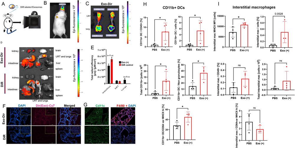 Figure 3. DiR labeling enabled visualization of exosome biodistribution in mice, showing respiratory tract and lung localization after intranasal administration, with further validation by organ imaging, lung tissue staining, and immune cell analysis. (Hui W W, et al., 2021)
- DiR-labeled exosomes were successfully visualized 24 hours after administration.
- Strong fluorescence signals were detected in the upper respiratory tract and lungs.
- Free DiR controls helped distinguish true exosome localization from dye background.
- Lung tissue analysis confirmed exosome accumulation and immune cell infiltration near pulmonary mucosa.
Conclusion
This case shows that optimized DiR labeling enables reliable in vivo tracking of exosome biodistribution and supports tissue-level validation of exosome delivery.
Looking to improve exosome tracking, imaging sensitivity, or biodistribution analysis? Creative Biostructure offers tailored exosome labeling solutions based on your detection modality, exosome source, and research goals. Contact us to start your project with a customized labeling strategy.
References
- Hui W W, Emerson L E, Clapp B, et al. Antigen-encapsulating host extracellular vesicles derived from Salmonella-infected cells stimulate pathogen-specific Th1-type responses in vivo. PLoS Pathogens. 2021, 17(5): e1009465.
- Santelices J, Ou M, Hui W W, et al. Fluorescent labeling of small extracellular vesicles (EVs) isolated from conditioned media. Bio-Protocol. 2022, 12(12): e4447-e4447.
- Boudna M, Campos A D, Vychytilova-Faltejskova P, et al. Strategies for labelling of exogenous and endogenous extracellular vesicles and their application for in vitro and in vivo functional studies. Cell Communication and Signaling. 2024, 22(1): 171.
- Hong S, Jo H C, Kim H L, et al. In vivo exosome imaging: applications of diverse visualization techniques. BMB Reports. 2025, 58(8): 340.
Frequently Asked Questions
For any inquiries, our support team is ready to help you get technical support for your research and maximize your experience with Creative Biostructure.